 "
"
File:HD-Fig 2-colicinE1 structure.png
From 2008.igem.org
Size of this preview: 800 × 284 pixels
Full resolution (1,502 × 534 pixels, file size: 382 KB, MIME type: image/png)
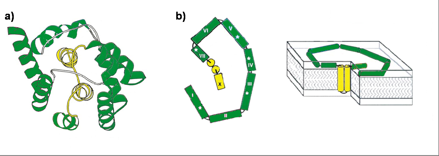
(a) Ribbon diagram of the Colicin E1 pore-forming domain by X ray crystal structure. The channel forming domain contains 10 helices (I-X). The two hydrophobic helices (VIII and IX) are labeled yellow. (b) Colicin E1 in a membrane bound state in two perspectives. Left panel shows the surface bound helices with an area of ~4200 Å2. Right panel shows colicin E1 bound to a lipid bilayer. The intra-membrane sections of the two helices VIII and IX are shown in yellow.
File history
Click on a date/time to view the file as it appeared at that time.
| Date/Time | Thumbnail | Dimensions | User | Comment | |
|---|---|---|---|---|---|
| current | 01:28, 29 October 2008 | 1,502×534 (382 KB) | Andreaskuehne (Talk | contribs) | ((a) Ribbon diagram of the Colicin E1 pore-forming domain by X ray crystal structure. The channel forming domain contains 10 helices (I-X). The two hydrophobic helices (VIII and IX) are labeled yellow. (b) Colicin E1 in a membrane bound state in two perspe) |
File links
The following 2 pages link to this file: