 "
"
File:Jr rmf 2.jpg
From 2008.igem.org
Size of this preview: 652 × 599 pixels
Full resolution (720 × 662 pixels, file size: 257 KB, MIME type: image/jpeg)
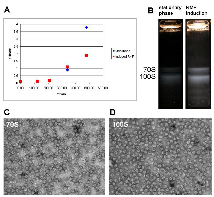
‘’’A’’’ Growth curve of BL21 DE3 cells holding the pET28a-RMF plasmid without induction (uninduced) and after induction after 5 hours. Cells were grown in 1l of medium in 5l baffled flasks (37C, 95rpm) and induced once they had reached the exponential phase. ‘’’B’’’ Ribosome profile of cells in the stationary phase and after RMF induction. Cells were harvested by centrifugation (SLC-6000 rotor, 7200rpm, 10min, 4C) and disrupted in a cell disruptor (Constant systems). The lysate was cleared at 13000 rpm in a SLA-1500 rotor in a Sorvall refrigerated centrifuge. Supernatant was collected and layered on top of a 30% sucrose solution (50mM HEPES-KOH pH 7.6; 100mM KCl; 10mM MgCl2; 1mM DTT). After centrifugation at 50000rpm for 20 hours at 4C in the preparative ultracentrifuge using a Ti70 rotor (Beckman Coulter) the supernatant was decanted and the pellet resuspended in ribosome buffer (50mM HEPES-KOH pH 7.6; 100mM KCl; 10mM MgCl2; 1mM DTT). The resuspended ribosomes were layered on top of a 10 to 40% (w/w) sucrose gradient, which was centrifuged in the SW32 swing rotor at 28000 rpm for 7 hours. Ribosome bands were visualized by light scattering (Tyndall effect) and photographed with a Nikon D80 SLR. ‘’’C’’’ and ‘’’D’’’ Electron micrographs of the 70S and 100S fraction show ribosomes in both fractions as the only constituent. Ribosome samples were diluted to OD260 = 1. 7ul of ribosome solution was added to a glow discharged carbon grid (Quantifoil) and stained with uranyl acetate according to standard protocol (1min sample adsorption, three times washing with 1% uranyl acetate solution by floatation). Samples were imaged with a FEI Morgagni 268 transmission electron microscope with tungsten emitter at an acceleration voltage of 100kV; 30000x magnification and recorded with a post column Gatan CCD camera.
File history
Click on a date/time to view the file as it appeared at that time.
| Date/Time | Thumbnail | Dimensions | User | Comment | |
|---|---|---|---|---|---|
| current | 23:41, 26 October 2008 | | 720×662 (257 KB) | Jrabl (Talk | contribs) | (‘’’A’’’ Growth curve of BL21 DE3 cells holding the pET28a-RMF plasmid without induction (uninduced) and after induction after 5 hours. Cells were grown in 1l of medium in 5l baffled flasks (37C, 95rpm) and induced once they had reached the exp) |
File links
The following page links to this file: