Team:Heidelberg/Human Practice/Open Day/Microscopy
From 2008.igem.org
(Difference between revisions)
(→Photo Gallery- Microscopy) |
(→Photo Gallery- Microscopy) |
||
| Line 4: | Line 4: | ||
{| align="center" | {| align="center" | ||
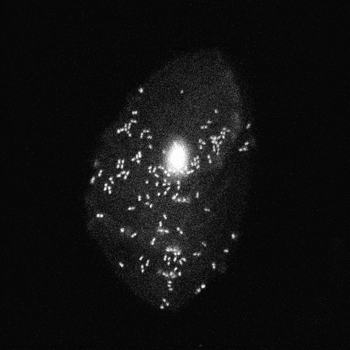
| - | | style="width:120px;" align="right" rowspan="3"| [[image:mchd_bac.gif|thumb|350 px| | + | | style="width:120px;" align="right" rowspan="3"| [[image:mchd_bac.gif|thumb|350 px| bacteria out of the mouth- group1_1]] |
| - | | style="width:120px;" align="right" rowspan="3"| [[image:mchd_Group1.gif|thumb|350 px| | + | | style="width:120px;" align="right" rowspan="3"| [[image:mchd_Group1.gif|thumb|350 px| bacteria out of the mouth- group1_2]] |
|} | |} | ||
{| align="center" | {| align="center" | ||
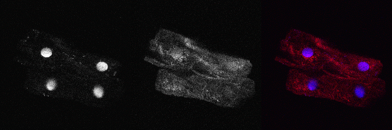
| - | | style="width:120px;" align="right" rowspan="3"| [[image:mchd_MontageGroup1.gif|thumb|350 px| | + | | style="width:120px;" align="right" rowspan="3"| [[image:mchd_MontageGroup1.gif|thumb|350 px| bacteria out of the mouth- staining- group1_1]] |
| - | | style="width:120px;" align="right" rowspan="3"| [[image:mchd_MontageGroup2.gif|thumb|350 px| | + | | style="width:120px;" align="right" rowspan="3"| [[image:mchd_MontageGroup2.gif|thumb|350 px| bacteria out of the mouth- staining- group1_2]] |
|} | |} | ||
{| align="center" | {| align="center" | ||
| - | | style="width:120px;" align="right" rowspan="3"| [[image:mchd_Group2.gif|thumb|350 px| | + | | style="width:120px;" align="right" rowspan="3"| [[image:mchd_Group2.gif|thumb|350 px| bacteria out of the mouth- group2_1]] |
| - | | style="width:120px;" align="right" rowspan="3"| [[image:mchd_Group3_1.gif|thumb|350 px| | + | | style="width:120px;" align="right" rowspan="3"| [[image:mchd_Group3_1.gif|thumb|350 px| bacteria out of the mouth- group2_2]] |
|} | |} | ||
{| align="center" | {| align="center" | ||
| - | | style="width:120px;" align="right" rowspan="3"| [[image:mchd_MontageGroup3_1.gif|thumb|350 px| | + | | style="width:120px;" align="right" rowspan="3"| [[image:mchd_MontageGroup3_1.gif|thumb|350 px| bacteria out of the mouth- staining- group2_1]] |
| - | | style="width:120px;" align="right" rowspan="3"| [[image:mchd_MontageGroup3_2.gif|thumb|350 px| | + | | style="width:120px;" align="right" rowspan="3"| [[image:mchd_MontageGroup3_2.gif|thumb|350 px| bacteria out of the mouth- staining- group2_2]] |
|} | |} | ||
{| align="center" | {| align="center" | ||
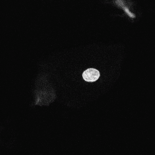
| - | | style="width:120px;" align="right" rowspan="3"| [[image:mchd_Mundschleimhaut.gif|thumb|350 px| | + | | style="width:120px;" align="right" rowspan="3"| [[image:mchd_Mundschleimhaut.gif|thumb|350 px| bacteria out of the mouth- group3_1]] |
| - | | style="width:120px;" align="right" rowspan="3"| [[image:mchd_Group3_2.gif|thumb|350 px| | + | | style="width:120px;" align="right" rowspan="3"| [[image:mchd_Group3_2.gif|thumb|350 px| bacteria out of the mouth- group3_2]] |
|} | |} | ||
{| align="center" | {| align="center" | ||
| - | | style="width:120px;" align="right" rowspan="3"| [[image:mchd_MontageMundschleimhaut.gif|thumb|350 px| | + | | style="width:120px;" align="right" rowspan="3"| [[image:mchd_MontageMundschleimhaut.gif|thumb|350 px| bacteria out of the mouth- staining- group3_1]] |
| - | | style="width:120px;" align="right" rowspan="3"| [[image:mchd_MontageBakterien.gif|thumb|350 px| | + | | style="width:120px;" align="right" rowspan="3"| [[image:mchd_MontageBakterien.gif|thumb|350 px| bacteria out of the mouth- staining- group3_2]] |
|} | |} | ||
Revision as of 02:34, 29 October 2008
Photo Gallery- Microscopy
Here you get an impression on what the pubils explored in the microscopy station.
 "
"


