Team:NYMU-Taipei/Project/Time Regulation/Results of Reloxilator
From 2008.igem.org
Blackrabbit (Talk | contribs) (→Results) |
Blackrabbit (Talk | contribs) (→Results) |
||
| (One intermediate revision not shown) | |||
| Line 54: | Line 54: | ||
Some of us say that the oscillations can be seen in that plot since the time period is around 46 minutes, however, some of us disagree. | Some of us say that the oscillations can be seen in that plot since the time period is around 46 minutes, however, some of us disagree. | ||
| + | |||
| + | |||
| + | [[Image:NYMU_RA3_LB.png]] | ||
| + | |||
| + | However what was weird was the plot of the LB. The fluorescence was higher than those that were measured in the other samples. | ||
| + | * Blue: 0 IPTG | ||
| + | * Pink: 50 IPTG | ||
==Reporting Assay 4== | ==Reporting Assay 4== | ||
| Line 67: | Line 74: | ||
** 470/530 | ** 470/530 | ||
===Results=== | ===Results=== | ||
| - | There was no significant difference between the wavelengths used. The shape looks the same to the human eye. But there was a very very weird trough between 0 to 2 hours. | + | There was no significant difference between the wavelengths used. The shape looks the same to the human eye. But there was a very very weird trough between 0 to 2 hours for all samples. |
Latest revision as of 05:04, 30 October 2008
Only the Oscillator, Tuner, and Reporter was successfully ligated together, so we performed a few Reporting Assays on it.
Contents |
Reporting Assay 1
This was done having little knowledge of what we were actually trying to look for. We just needed some data to get a feel for what kind of information we were looking for, and we also used this experiment as an indicator to how much time the experiment would take and what we could improve upon next time (e.g. how to take the samples from the tubes and put it back in the incubator in the shortest amount of time in order not to affect the experiment too much).
Method
Diluted all samples by a 1:100 ratio and let them grow. Measured the OD and fluorescence every hour.
Results
- Found that doing technical replicates actually makes a lot of difference if you didn't pipette properly.
- If starting with and OD600 of around 0.0325, the fast growth period where we see the most activity lies between 2 to 6 hours. From the next experiment on, we decided to dilute all samples to start off with an OD around 0.0325.
- Other than that, at first we had thought that the fluorescence values were too close to the blank (LB medium), and some even lower than it, so we did not analyse the data. It wasn't until after the third reporting assay we noticed that the measured points did indeed have a sinusoidal shape.
Reporting Assay 2
Method
Diluted the samples to start off with an OD600 of 0.0325. Measured the OD600 and a few different excitation/emission wavelengths.
Reporting Assay 3
Hogged the fluorescence measuring machine for 4 hours to let it grow in the machine. Doing this we could measure the fluorescence every five minutes. However, we couldn't measure the OD600.
Method
Samples consisted of:
- LB
- 0uM IPTG
- 50uM IPTG
- Oscillator + Reporter
- 0uM IPTG
- 50uM IPTG
- Oscillator + Tuner + Reporter
- 0uM IPTG
- 50uM IPTG
- 500uM IPTG
- 1000uM IPTG
We measured the samples every 5 minutes during the time period between 2 to 6 hours from the time of dilution.
Results
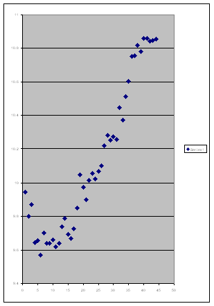
By this point, we were thinking about what we should do about getting "no results" every time since everytime we saw that the fluoresence of the blank sample was higher or similar to the fluoresence of the other samples. Then we plotted of of the samples by itself without the blank and alas! we saw a sinusoidal shape.
And our hope was revived! We grouped similar samples together and plotted them.
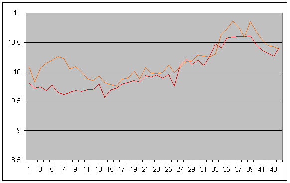
This is the plot of the oscillators + reporter. (each timestep is 5 minutes)
- Red: 0 IPTG
- Orange: 50 IPTG
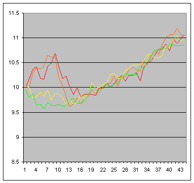
This is the plot of the oscillators + tuner + reporter. (each timestep is 5 minutes)
- Red: 0uM IPTG
- Orange: 50uM IPTG
- Yellow: 500uM IPTG
- Green: 1000uM IPTG
Some of us say that the oscillations can be seen in that plot since the time period is around 46 minutes, however, some of us disagree.
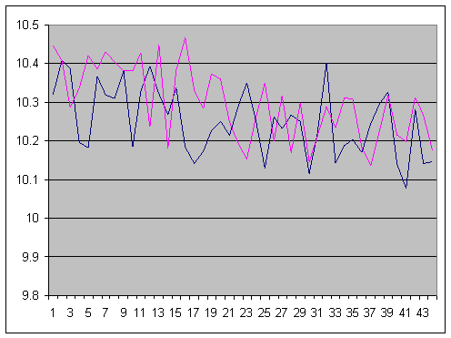
However what was weird was the plot of the LB. The fluorescence was higher than those that were measured in the other samples.
- Blue: 0 IPTG
- Pink: 50 IPTG
Reporting Assay 4
Started doing reporting assay of the Oscillator+Tuner+Synchronizer+Reporter construct, until we found out halfway that the construct had actually failed (we were using the time between measurement assay points to validate the length of the part from plasmid extraction->digestion->gel electrophoresis.
Reporting Assay 5
- Aim
- purely wanted to see the relationship that different amounts of IPTG has upon the fluorescence of the Oscillator+Tuner+Reporter construct. So OD600 was not measured except at the start.
- IPTG concentrations: 0,50,100,200,300,500,1000,2000,4000 uM.
- Measurement: every 5 minutes at the wavelengths (ex/em):
- 488/533
- 488/525
- 475/515
- 470/530
Results
There was no significant difference between the wavelengths used. The shape looks the same to the human eye. But there was a very very weird trough between 0 to 2 hours for all samples.
 "
"


