Team:Chiba/Calendar-Home/18 October 2008
From 2008.igem.org
(Difference between revisions)
(→results) |
|||
| (6 intermediate revisions not shown) | |||
| Line 3: | Line 3: | ||
[[Team:Chiba/Calendar-Home/17 October 2008|17 October 2008 <]]|[[Team:Chiba/Calendar-Home/19 October 2008|> 19 October 2008]] | [[Team:Chiba/Calendar-Home/17 October 2008|17 October 2008 <]]|[[Team:Chiba/Calendar-Home/19 October 2008|> 19 October 2008]] | ||
| + | ==Laboratory work== | ||
===Team:Demo-Rs=== | ===Team:Demo-Rs=== | ||
17 October~ | 17 October~ | ||
| Line 19: | Line 20: | ||
[[Image:Team-Chiba-P1000911.JPG|150px]] | [[Image:Team-Chiba-P1000911.JPG|150px]] | ||
[[Image:Team-Chiba-P1000915.JPG|150px]] | [[Image:Team-Chiba-P1000915.JPG|150px]] | ||
| - | [[Image:Team-Chiba-P1000923.JPG| | + | [[Image:Team-Chiba-P1000923.JPG|145px]] |
| + | [[Image:Team-Chiba-P1000931.JPG|143px]] | ||
| + | [[Image:Team-Chiba-P1000936.JPG|140px]] | ||
| + | 0h 0.5h 1.0h 1.5h 2.0h | ||
Latest revision as of 00:40, 30 October 2008
17 October 2008 <|> 19 October 2008
Laboratory work
Team:Demo-Rs
17 October~
- Sender Wash
- Centrifuged 2 tubes containing([http://partsregistry.org/Part:BBa_T9002 BBa_T9002](JW1908))at 20°C,3600rpm for 6min and discarded supernatant.
- Added 10mL LB-Amp to each tube.
- Repeated wash twice.
- Creating bacterial plates
- LB-Amp pre-cultured Sender([http://partsregistry.org/Part:BBa_S03623 BBa_S03623](JW1908)) tube 1 (10mL) was mixed with LB-Amp-agar(50°C)(10ml)to produce sender containing bacterialplate-1.
- Lifted with nitrocellulose
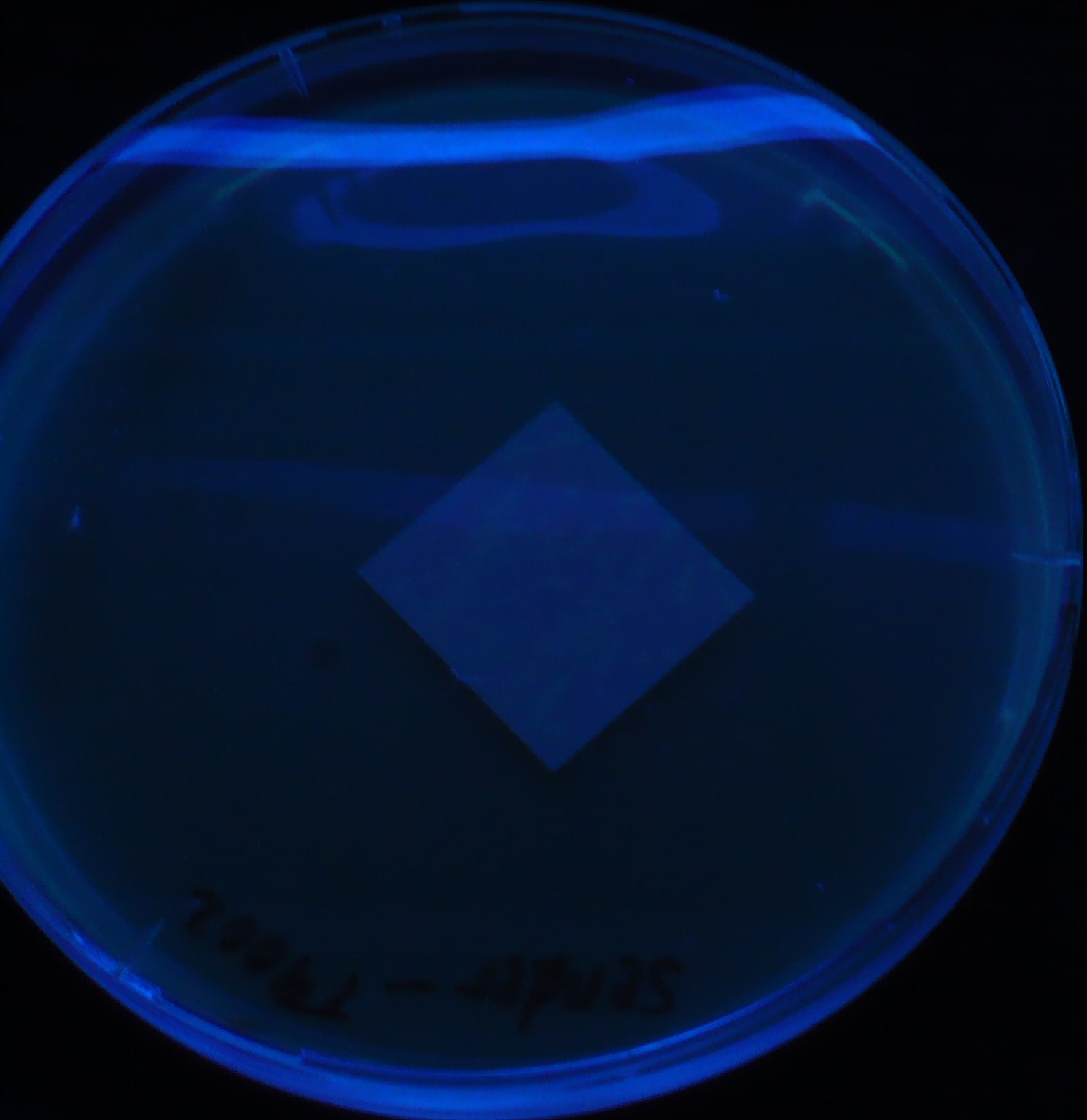
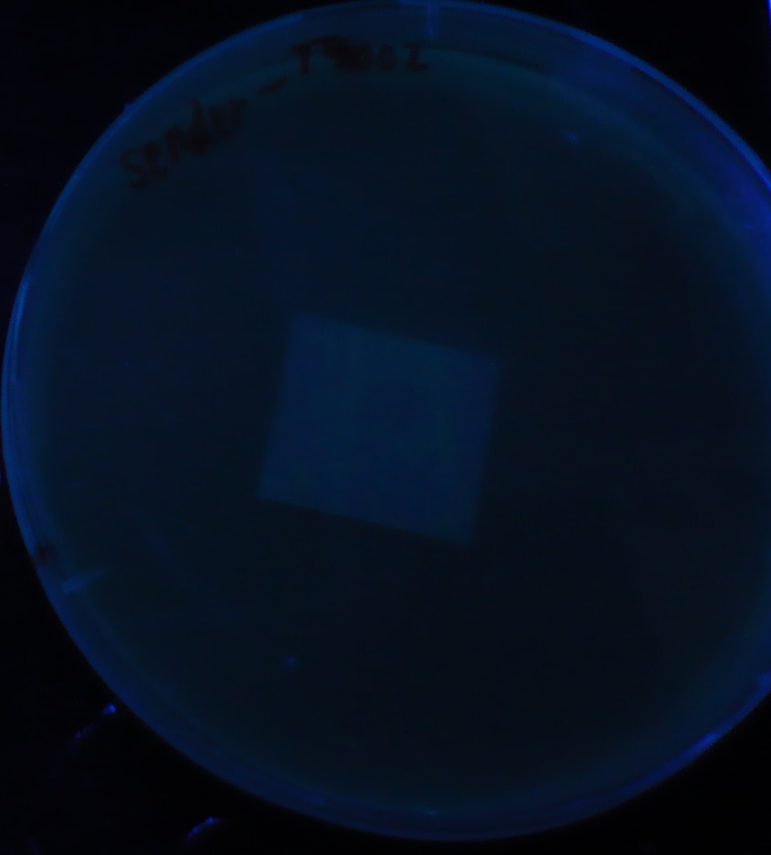
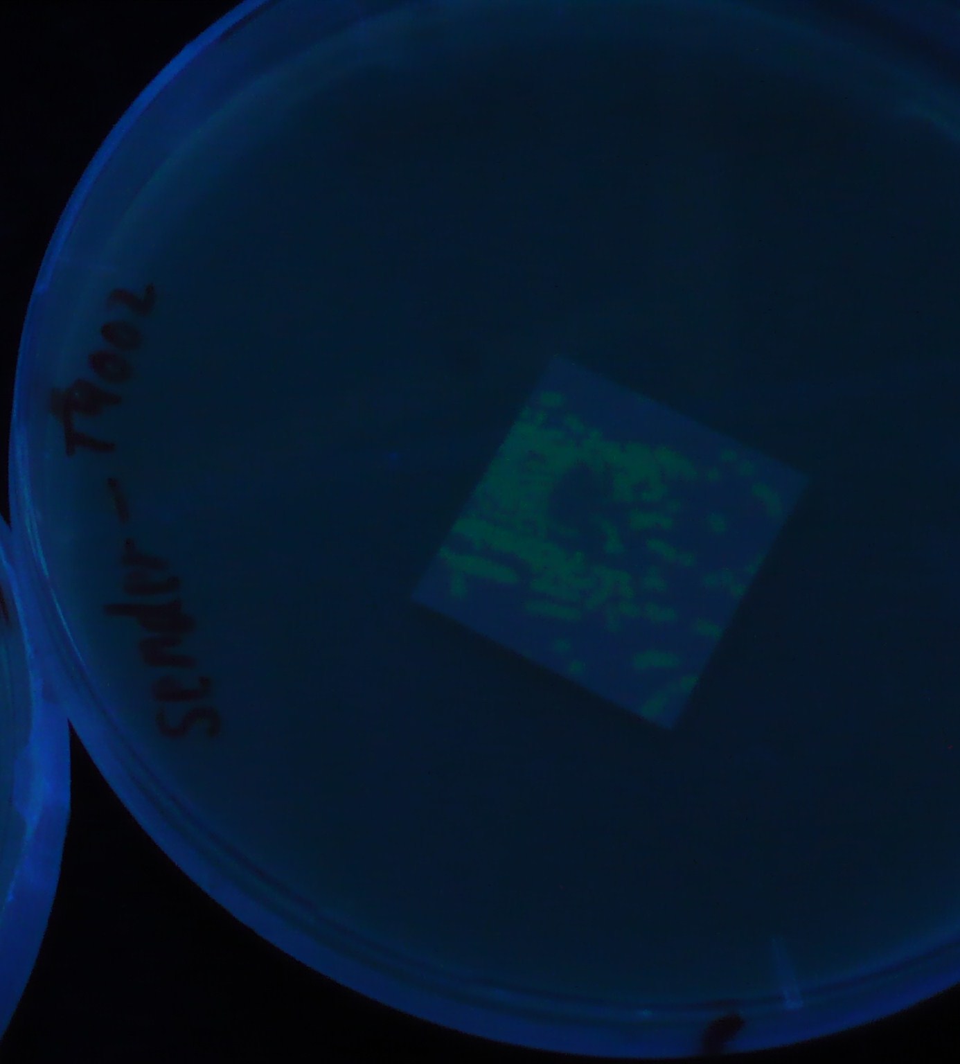
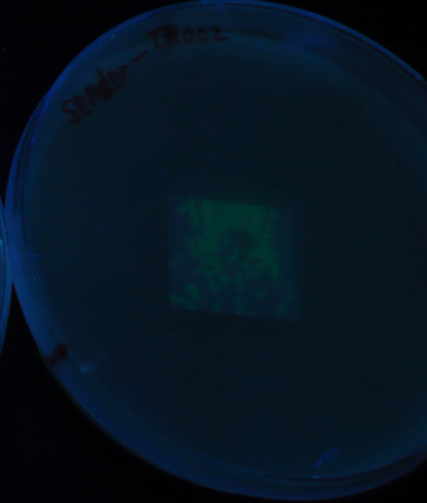
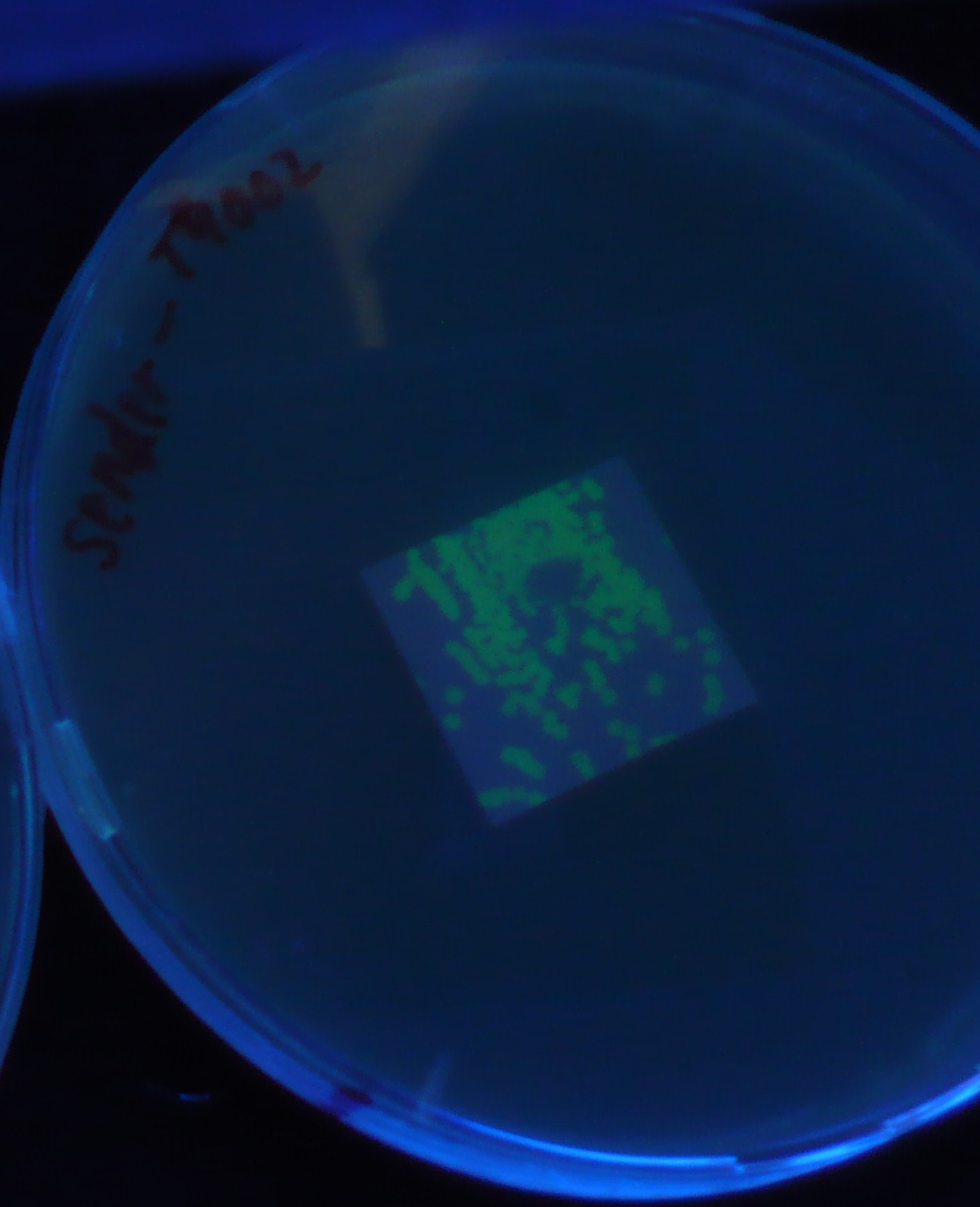
- Receiver([http://partsregistry.org/Part:BBa_T9002 BBa_T9002](JW1908))colony was transfered to a nitrocellulose filter and placed on each of Sender([http://partsregistry.org/Part:BBa_S03623 BBa_S03623](JW1908))containing bacterial plate (1~3) and Sender-absent negative control plate (t=0). Determined the time required for the colonies to fluoresce depending on the bacterial concentration (100 and 1000-fold dilution).
- Method to detect fluorescence
- Plates cultured at 37°C were exposed to UV (312nm) light once every 30 minutes to observe GFP fluorescence.
results
0h 0.5h 1.0h 1.5h 2.0h
 "
"
