Team:Freiburg Transfection
From 2008.igem.org
(Difference between revisions)
| Line 3: | Line 3: | ||
<div style="font-size:18pt;"> | <div style="font-size:18pt;"> | ||
<font face="Arial Rounded MT Bold" style="color:#010369">_Transfection</font></div> | <font face="Arial Rounded MT Bold" style="color:#010369">_Transfection</font></div> | ||
| - | |||
<br> | <br> | ||
<h3>'''Testing the transfection protocol'''</h3> | <h3>'''Testing the transfection protocol'''</h3> | ||
| Line 10: | Line 9: | ||
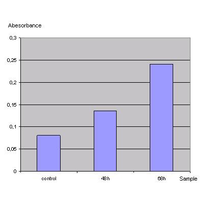
ONPG-assay: <br> | ONPG-assay: <br> | ||
<br>[[Image:FreiGEMTable3onpg.JPG]]<br> | <br>[[Image:FreiGEMTable3onpg.JPG]]<br> | ||
| - | '' | + | ''Table 1_Transfection: Absorbance of o-Nitrophenol produced by the β-galactosidase'' |
| - | + | <br> | |
[[Image:Graph2onpg.JPG]]<br> | [[Image:Graph2onpg.JPG]]<br> | ||
| - | '' | + | ''Graph 1_Transfection: Absorbance of o-Nitrophenol produced by the β-galactosidase''<br> |
Control was done with untransfected cells using the same procedure.<br> | Control was done with untransfected cells using the same procedure.<br> | ||
<br> | <br> | ||
| Line 21: | Line 20: | ||
<br> | <br> | ||
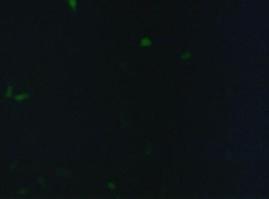
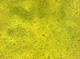
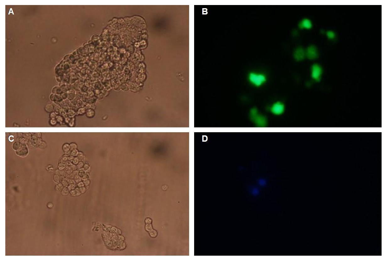
[[Image:Freiburg2008_150%_293Tzvi.jpg]] [[Image:Freiburg2008_Kontrolle.jpg]] [[Image:Freiburg2008_Kontrolle_Durchlicht.jpg]]<br> | [[Image:Freiburg2008_150%_293Tzvi.jpg]] [[Image:Freiburg2008_Kontrolle.jpg]] [[Image:Freiburg2008_Kontrolle_Durchlicht.jpg]]<br> | ||
| + | ''Figure 1_Transfection''<br> | ||
<br> | <br> | ||
| Line 26: | Line 26: | ||
<h3>'''Localization at the cell membrane'''</h3> | <h3>'''Localization at the cell membrane'''</h3> | ||
To show the localization of the constructs at the cell membrane transfection of the construct signalpeptide-Lipocalin-transmembraneregion-betaLactamase1-YFP was performed.<br> | To show the localization of the constructs at the cell membrane transfection of the construct signalpeptide-Lipocalin-transmembraneregion-betaLactamase1-YFP was performed.<br> | ||
| - | Figure | + | Figure 2_Transfection shows the configuration of the construct. Lipocalin, the fluorescein binding Anticalin, exhibits the extracellular part of the construct. The transmembrane region is appropriate to that of the EGF-receptor erbb1. Split-beta-Lactamase, the intracellular part is labeled to the yellow fluorescent protein to detect membrane localization.<br> |
<br> | <br> | ||
[[Image:Freiburg2008_Lipo_bla1+YFP.jpg|500px]] | [[Image:Freiburg2008_Lipo_bla1+YFP.jpg|500px]] | ||
<br> | <br> | ||
| - | '''Figure | + | '''Figure 2_Transfection''' |
<br> | <br> | ||
<br> | <br> | ||
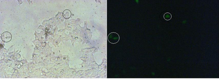
| - | Membranelocalization of the construct signalpeptide-Lipocalin-transmembraneregion-betaLactamase1-YFP is visible in transfected 293T cells (Figure | + | Membranelocalization of the construct signalpeptide-Lipocalin-transmembraneregion-betaLactamase1-YFP is visible in transfected 293T cells (Figure 3_Transfection). The fluorescence of the cells is most likely restricted to the cellmembrane which confirms the assembly of the construct in the cytoplasmamembrane.<br> |
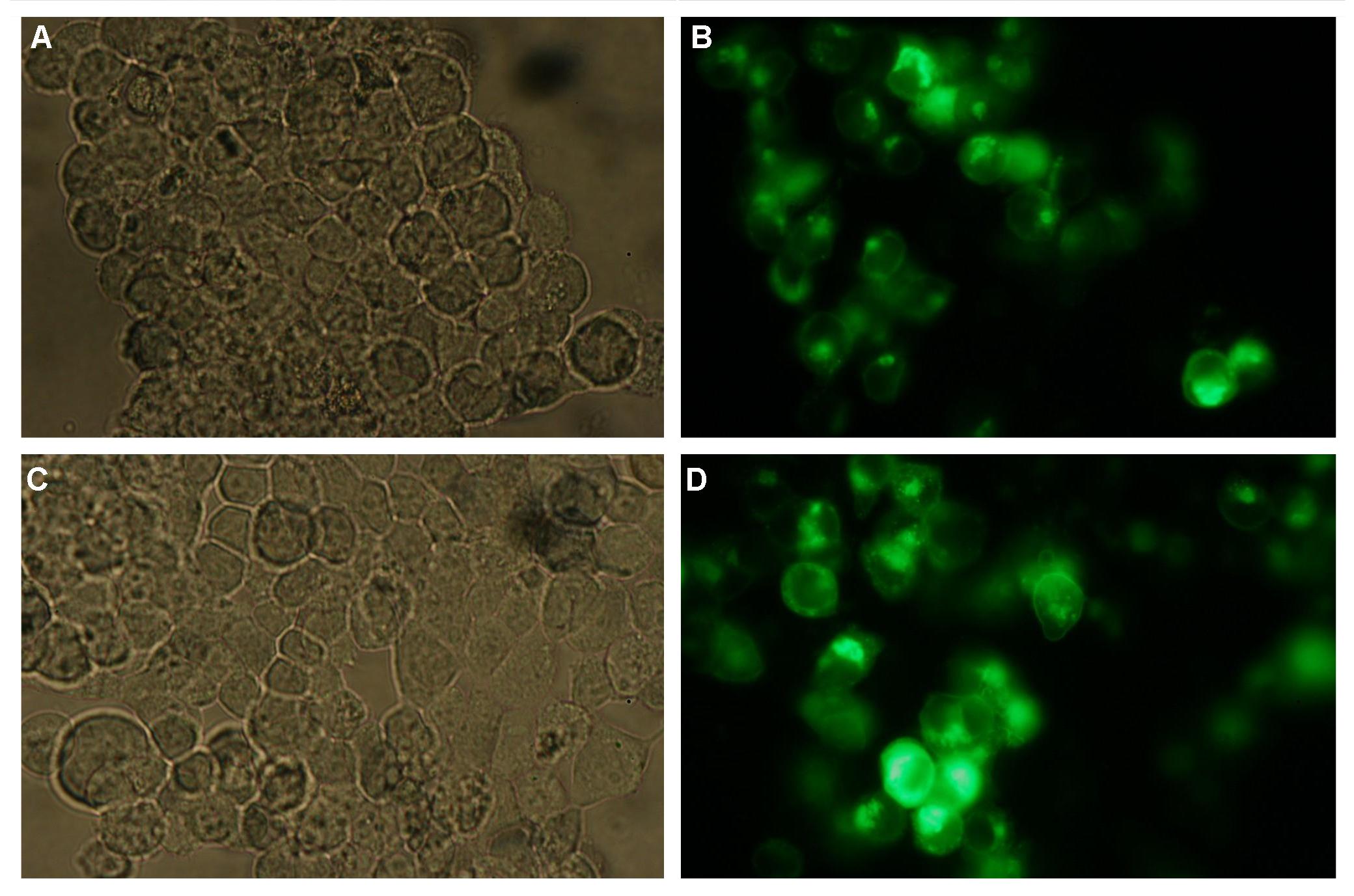
| - | In comparison, 293T cells transfected with the construct transfectionvector-YFP show a uniformly distributed fluorescence all-over the cell (Figure | + | In comparison, 293T cells transfected with the construct transfectionvector-YFP show a uniformly distributed fluorescence all-over the cell (Figure 4_Tansfection A and B).<br> |
| - | Transfection with the construct transfectionvector-CFP as well results in completely fluorescent cells (Figure | + | Transfection with the construct transfectionvector-CFP as well results in completely fluorescent cells (Figure 4_Transfection C and D).<br> |
<br> | <br> | ||
[[Image:Freiburg2008_SP_LIPO_GGGS_TM_bla1_YFP_1.jpg|710px]]<br> | [[Image:Freiburg2008_SP_LIPO_GGGS_TM_bla1_YFP_1.jpg|710px]]<br> | ||
| - | '''Figure | + | '''Figure 3_Transfection'''<br> |
<br> | <br> | ||
[[Image:Freiburg2008_TV_CMV_YFP___CFP_loeslich.jpg|700px]]<br> | [[Image:Freiburg2008_TV_CMV_YFP___CFP_loeslich.jpg|700px]]<br> | ||
| - | '''Figure | + | '''Figure 4_Transfection'''<br> |
<br> | <br> | ||
<h3>'''Double transfections with Splitfluorophor-/Splitenzyme-constructs'''</h3> | <h3>'''Double transfections with Splitfluorophor-/Splitenzyme-constructs'''</h3> | ||
| - | On Figure | + | On Figure 5_Transfection the structures of the signalpeptide-Lipocalin-transmembraneregion-nCFP and signalpeptide-Lipocalin-transmembraneregion-fluolinker-cCFP are visible (exemplary for the Splitfluorophore-/Splitenzyme-constructs). The extracellular fragment is build of Lipocalin (fluorescein binding Anticalin) and a GGGSLinker. Intracellular either the N-terminal part or the C-terminal part of the splitfluorophore is fused to the transmembrane region of the EGF-receptor. To achieve more flexibility and to support the assembly of the two splitfluorophore parts a fluolinker is fused in between the transmembrane region and the C-terminal part of the splitfluorophores.<br> |
<br> | <br> | ||
[[Image:Freiburg2008_Lipo+Split_CFP.jpg|450px]]<br> | [[Image:Freiburg2008_Lipo+Split_CFP.jpg|450px]]<br> | ||
| - | '''Figure | + | '''Figure 5_Transfection'''<br> |
<br> | <br> | ||
Adding fluorescein-coupled molecules leads to a clustering of the Lipocalin constructs due to the fluorescein-Lipocalin-binding (Similarly Nip-coupled molecules lead to a clustering of Nip constructs). | Adding fluorescein-coupled molecules leads to a clustering of the Lipocalin constructs due to the fluorescein-Lipocalin-binding (Similarly Nip-coupled molecules lead to a clustering of Nip constructs). | ||
| - | The clustering of the constructs in turn results in an assembly of the splitfluorophores or splitenzymes and therefore creates a functional protein (Figure | + | The clustering of the constructs in turn results in an assembly of the splitfluorophores or splitenzymes and therefore creates a functional protein (Figure 6_Transfection).<br> |
<br> | <br> | ||
[[Image:Freiburg2008_Lipo+Split_YFP.jpg|450px]]<br> | [[Image:Freiburg2008_Lipo+Split_YFP.jpg|450px]]<br> | ||
| - | '''Figure | + | '''Figure 6_Transfection'''<br> |
<br> | <br> | ||
Revision as of 17:21, 28 October 2008
 "
"


