Preparing Bacillus subtilis for Microscopy
From 2008.igem.org
(Difference between revisions)
Riachalder (Talk | contribs) |
Riachalder (Talk | contribs) |
||
| Line 3: | Line 3: | ||
To view ''B. subtilis'' cells under the microscope, cells must first be immobilised in order to allow the long exposure time to give a clear picture. | To view ''B. subtilis'' cells under the microscope, cells must first be immobilised in order to allow the long exposure time to give a clear picture. | ||
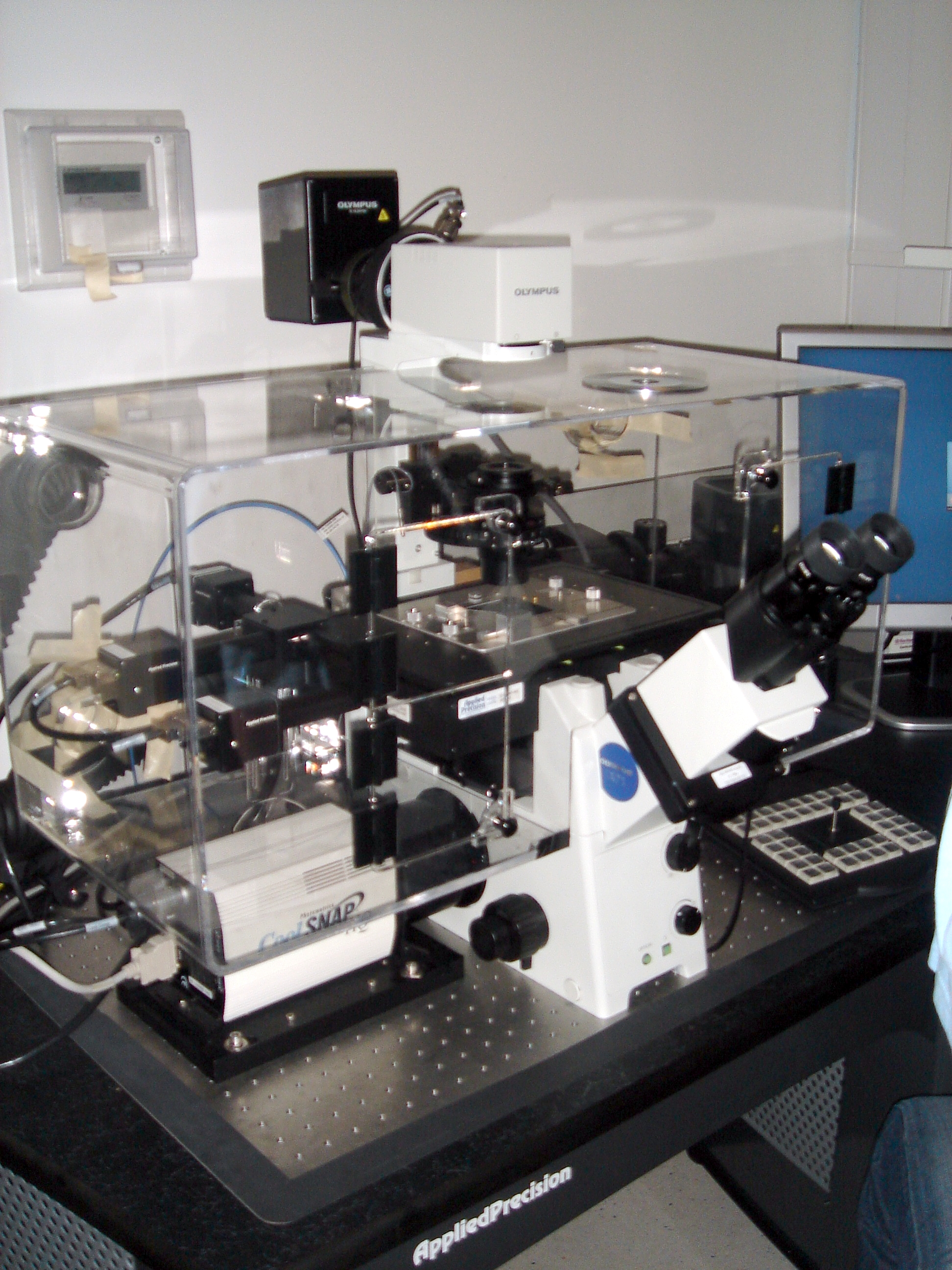
[[Image:DSC01655.jpg|thumb|left|200px|Microscope]] | [[Image:DSC01655.jpg|thumb|left|200px|Microscope]] | ||
| - | |||
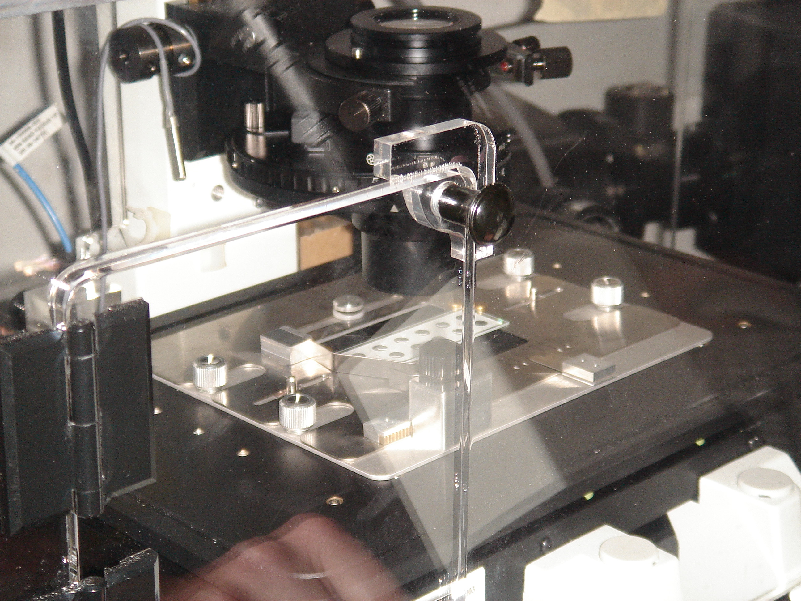
To do this, make a solution of 1g agarose in 100mL SMM and micorwave to dissolve the solute. (This can then be kept liquid in an oven for future use.) Place droplets of this in the wells of a microscope slide (opne droplet for each sample to be viewed) and leave for 1-2 minutes until the gel has almost set. Place samples on the droplets and press a coverslip on firmly. | To do this, make a solution of 1g agarose in 100mL SMM and micorwave to dissolve the solute. (This can then be kept liquid in an oven for future use.) Place droplets of this in the wells of a microscope slide (opne droplet for each sample to be viewed) and leave for 1-2 minutes until the gel has almost set. Place samples on the droplets and press a coverslip on firmly. | ||
| + | [[Image:DSC01656.jpg|thumb|center|200px]] | ||
Revision as of 10:28, 15 September 2008
To view B. subtilis cells under the microscope, cells must first be immobilised in order to allow the long exposure time to give a clear picture.
To do this, make a solution of 1g agarose in 100mL SMM and micorwave to dissolve the solute. (This can then be kept liquid in an oven for future use.) Place droplets of this in the wells of a microscope slide (opne droplet for each sample to be viewed) and leave for 1-2 minutes until the gel has almost set. Place samples on the droplets and press a coverslip on firmly.
 "
"


