Team:NTU-Singapore/Parts/Characterization of pLsrA-YFP
From 2008.igem.org
(New page: <html><link rel="stylesheet" href="http://greenbear88.googlepages.com/ntu_igem.css" type="text/css"></html> <div id="header">{{User:Greenbear/sandbox/header}}</div> <div id="mainconten...)
Newer edit →
Revision as of 01:15, 24 October 2008
|
Characterization of plsrA-YFP
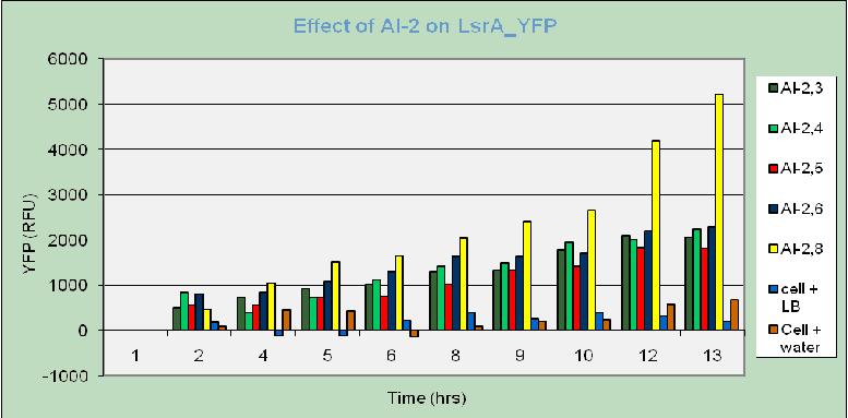
For all the samples with AI-2 added, relative fluorescence unit (RFU) increases with time. In contrast, the cell samples with 50 µl water added have their RFU low and not varied much. The control samples with addition of 50 µl of LB have higher RFU level, yet it remained steady with time. From this observation, it can be concluded that the increase in RFU of the samples with AI-2 addition is not related to cell growth as as result of LB presence in the supernatants. Hence, this increasing trend can be well explained as YFP was actually expressed in the presence of AI-2. This further proves that the LsrA promoter part BBa_K117002 in fact works correctly as it is activated in the presence of AI-2. Further characterization of this promoter will be implemented in future works. From this result, it is highly expected that the detection system part BBa_K117010 also expresses lysis protein (celE7) upon induced by AI-2.
This clustered column graph shows a clearer view on RFU expressed by different samples throughout the experiment. As expected, RFU levels of the control samples (cell suspension with addition of water and LB) are very low and not varied much compared to the high and increasing RFU of the samples with AI-2-containing supernatants.
Another conclusion can be drawn is that AI-2 activity seems to be highest for the supernatants obtained after 8 hours of incubation. "
"


