Team:NYMU-Taipei/Project/Urea
From 2008.igem.org
Blackrabbit (Talk | contribs) |
Blackrabbit (Talk | contribs) |
||
| Line 2: | Line 2: | ||
== Why should urea be removed == | == Why should urea be removed == | ||
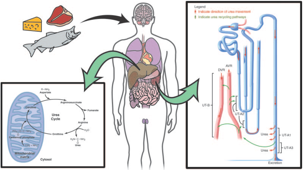
| - | Physiologically, urea is synthesized from ammonia and carbon dioxide. The elimination of urea means getting rid of nitrogenous waste from the body. In healthy individuals, blood plasma | + | Physiologically, urea is synthesized from ammonia and carbon dioxide. The elimination of urea means getting rid of nitrogenous waste from the body. In healthy individuals, blood plasma are filtrated by kidney and the waste removed by urination; in patients with renal failure, such waste are accumulated in blood. These patients have to get renal dialysis treatment to remove the waste, or they would suffer from hepatic failure and encephalopathy, which is worse. For these reasons, urea should be removed by a healthy kidney or by medical treatment. |
However, the renal dialysis treatment knocks down the quality of life because of the time spent being treated and equipment portage (or patient transportation). If urea can be removed by the enterobacteria (bacteria in intestines, such as ''E. coli'' or our BacToKidney), a capsule or an yogurt-like drug may help reduce the frequency of get renal dialyzed and increase the quality of life. If so, the next half of the patients' lifespan may be more fun. | However, the renal dialysis treatment knocks down the quality of life because of the time spent being treated and equipment portage (or patient transportation). If urea can be removed by the enterobacteria (bacteria in intestines, such as ''E. coli'' or our BacToKidney), a capsule or an yogurt-like drug may help reduce the frequency of get renal dialyzed and increase the quality of life. If so, the next half of the patients' lifespan may be more fun. | ||
| + | |||
| + | [[Image:NYMU_urea transport_in_human_body.png|800px]] | ||
== How do we try to remove urea == | == How do we try to remove urea == | ||
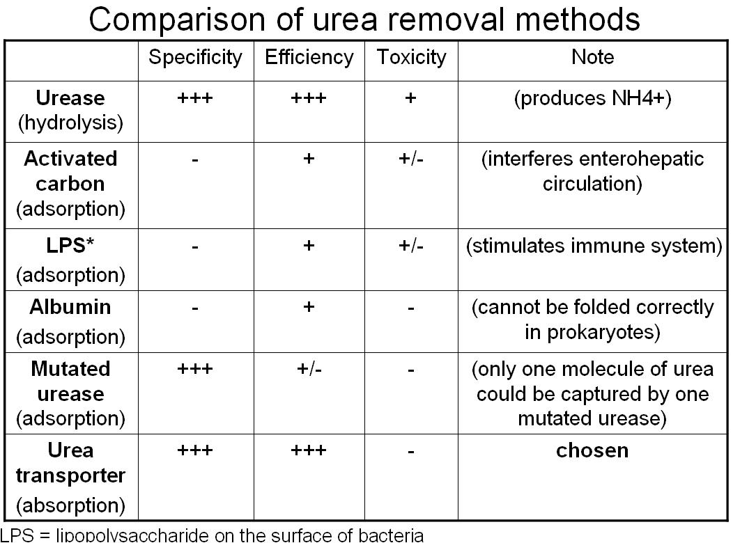
| + | [[Image:NYMU_urea_CompareUreaRemovalMethods.jpg|600px]] | ||
| - | |||
| + | |||
| + | === Absorption Method--Urea Transporter (chosen method) === | ||
| + | |||
| + | *Urea may be absorbed by the micro dialysis machine (or be uptaken by the engineered bacteria), and this micro machine may be eliminated with stool passage. | ||
| + | |||
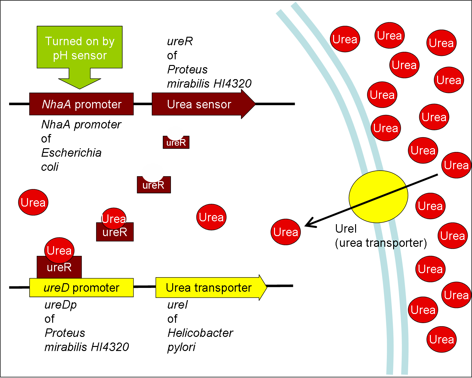
| + | *Urea transporter would be expressed only if both pH sensor and urea sensor are turned on: | ||
| + | |||
| + | *When the device is turned on by pH sensor, ureR, the pH sensor is expressed. The ureR protein may bind with urea molecule to form a complex. This ureR-urea complex may activate a downstream promoter, ureDp, and then urea transporter will be expressed. | ||
[[Image:NYMU_Urea0731.png|600px]] | [[Image:NYMU_Urea0731.png|600px]] | ||
| + | |||
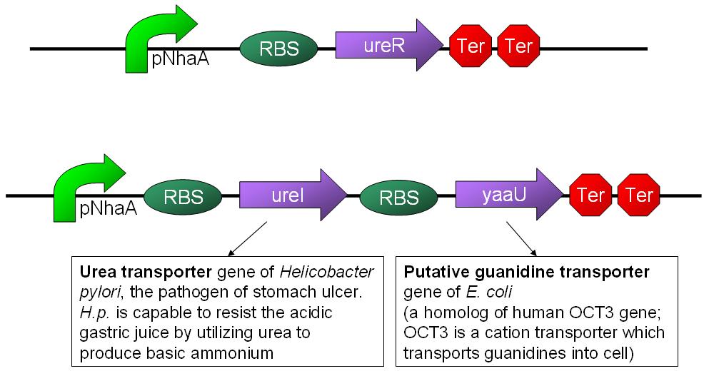
| + | *In this circuit design, urea sensor turns not only urea transporter on but also guanidine transporter on at the same time. Guanidine transporter is integrated into this circuit because the concentratoin of guanidine in blood may be elevated after an rising of urea consentration. | ||
| + | [[Image:NYMU_urea_biobrick.jpg|600px]] | ||
=== Adsorption Method === | === Adsorption Method === | ||
| Line 25: | Line 38: | ||
== How to measure the concentration of urea == | == How to measure the concentration of urea == | ||
| - | + | ===== Urease method ===== | |
| + | Urea + H2O --(urease)--> 2NH4+ + CO2 | ||
| + | NH4+ + alpha-KG + NADH --(GLDH)--> Glutamate + NAD+ + H2O | ||
| + | |||
| + | The change of NADH concentration reflects the urea concentration in the solution. | ||
| + | |||
| + | Absorbance of NADH = A340nm - A405nm | ||
| + | |||
| + | |||
| + | * use an in vitro diagnostic kit | ||
| + | ** [http://www.fbc.com.tw/j2fbs/fbc_tw/medical/24-pdf/BUN_dream%201.0.pdf Formosa Biomedical Technology] Blood Urea Nitrogen diagnostic kit | ||
| + | * or undertaken by clinical laboratories | ||
| + | ** [http://www.ucl.com.tw/WebMaster/?section=92 UCL (Union Clinical Laboratory)] A test for urea nitrogen costs NTD$60 (as private patient without health insurance) | ||
| + | |||
| + | ===== Diacetyl monoxime method: ===== | ||
Diacetyl Monoxime + Urea --> Diazine (yellow) | Diacetyl Monoxime + Urea --> Diazine (yellow) | ||
| Line 32: | Line 59: | ||
* [http://www.clinchem.org/cgi/content/abstract/15/5/393 The Automated Thiosemicarbazide-Diacetyl Monoxime Method for Plasma Urea, Clinical Chemistry 15: 393-396, 1969] | * [http://www.clinchem.org/cgi/content/abstract/15/5/393 The Automated Thiosemicarbazide-Diacetyl Monoxime Method for Plasma Urea, Clinical Chemistry 15: 393-396, 1969] | ||
{{:Team:NYMU-Taipei/Footer}} | {{:Team:NYMU-Taipei/Footer}} | ||
| + | |||
| + | == Expieriments == | ||
| + | |||
| + | {| border="1" | ||
| + | |- | ||
| + | | <font color="green">'''Measurement of Urea'''</font> || | ||
| + | |- | ||
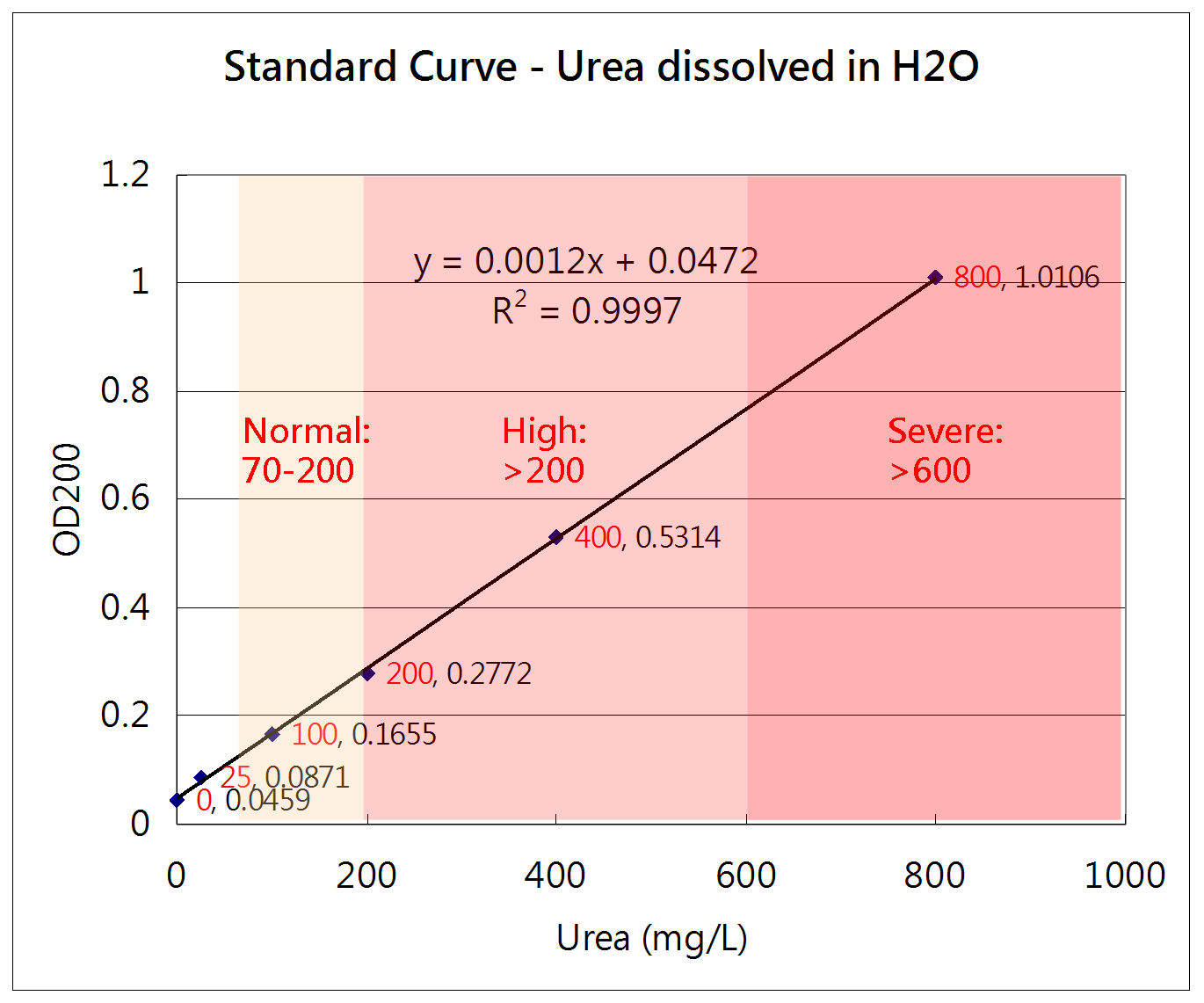
| + | | [[Image:NYMU_urea_exp_0821.png|400px]] || | ||
| + | *08/21, Ming-Han & Blent | ||
| + | *Urea can be dissolved in ddH2O, and 8000mg/L stock has been prepared. | ||
| + | *The Absorbance (200nm) of urea dissolved in ddH2O shows good linearity in a range from 0 to 800 mg/L. | ||
| + | *The normal range of urea concentration in blood is 70-200mg/L | ||
| + | *Traditional renal dialysis could take urea 50-75% off blood | ||
| + | |- | ||
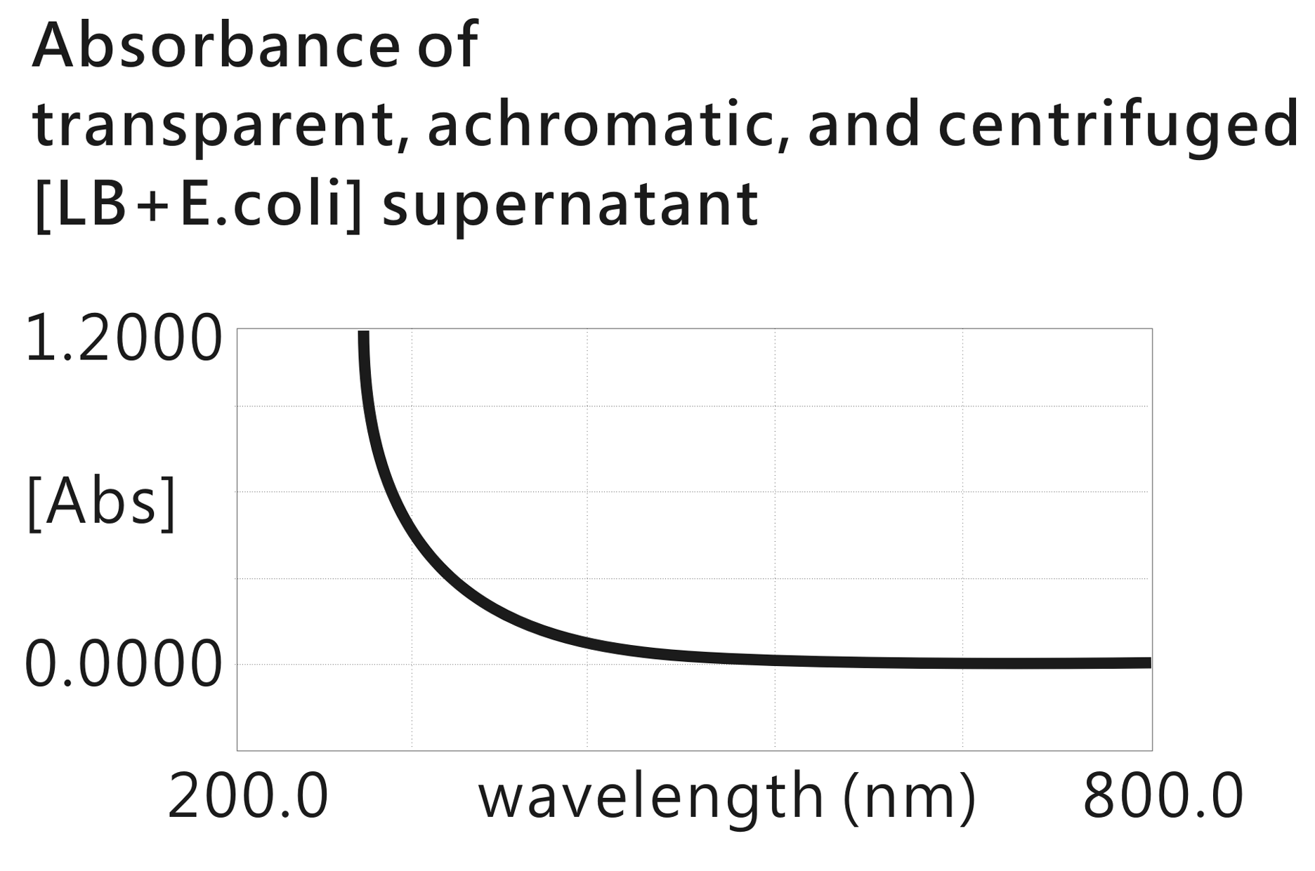
| + | | [[Image:NYMU_urea_exp_0822.png|400px]] || | ||
| + | *08/22, Ming-Han | ||
| + | *There is something that may interfere with measuring OD200 greatly existing in the ''E.coli''-grown LB broth. | ||
| + | *The result implies the OD200 method cannot help calculating the urea quantity removed by our engineered bacteria. Orz | ||
| + | |- | ||
| + | | | ||
| + | [[Image:NYMU_uera_in_LB.PNG|400px]] | ||
| + | * BUN = Blood Urea Nitrogen | ||
| + | * Urea conc. of LB 0.1X is <1 mg/dL (<10 mg/L) | ||
| + | * Urea conc. of LB 0.5X is 1.4 mg/dL (=14 mg/L) | ||
| + | * '''Urea conc. of LB 1X is 2.1 mg/dL (=21 mg/L)''' | ||
| + | * Urea conc. of blood is 7-20 mg/dL (=70-200 mg/L) | ||
| + | || | ||
| + | * 08/28, Ming-Han & UCL (Union Clinical Laboratory) | ||
| + | * Conclusion: LB is a kind of competent medium for quantification of urea, because its internal urea concentration (21 mg/L) is much lower than the blood urea concentration both in health individuals (70-200 mg/L) and nephropathic patients (>200 mg/L). | ||
| + | * Potential drawback: the urea contained in LB might interfere the measurement of the urea removal ability of our engineered bacteria if (1) the contents of LB varies from a batch to a batch (2) our urea scavenger bacteria make little significance. | ||
| + | |- | ||
| + | | '''<font color="green">Construction</font>''' || | ||
| + | |- | ||
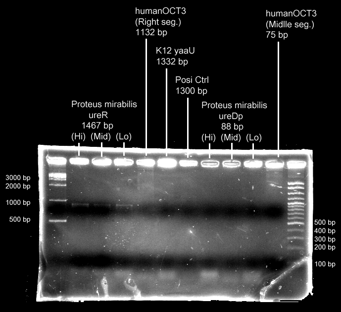
| + | | [[Image:NYMU_G_and_U_PCR20080904.png|400px]] || | ||
| + | * 09/05, Ming-Han & Yuan-Hao | ||
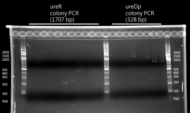
| + | * ureR and ureDp are (successfully) amplified. | ||
| + | * Program "G and U" in Little White, the PCR machine. | ||
| + | |- | ||
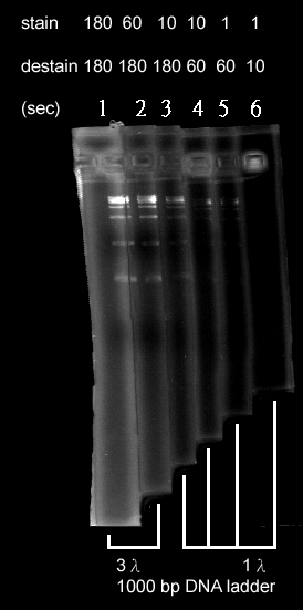
| + | | [[Image:NYMU_EtBr_test_labeled.png|150px]] || | ||
| + | * 09/07, Ming-Han | ||
| + | * Lane 1 = stain 3', destain 3' | ||
| + | * Lane 2 = stain 1', destain 3' | ||
| + | * Lane 3 = stain 10", destain 3 | ||
| + | * Lane 4 = stain 10", destain 1' | ||
| + | * Lane 5 = stain 1", destain 1" | ||
| + | * Lane 6 = stain 1", destain 10" | ||
| + | * Discussion/Conclusion: | ||
| + | ** The best way is to stain 3 or 1 min and then destain 3 min, when using the latest prepared EtBr solution. | ||
| + | ** To stain for 3 minutes might cause the whole gel brightened when using softer agarose gel. | ||
| + | ** The time of destaining is also important; maybe this step could be considered as fine staining. | ||
| + | |- | ||
| + | | | ||
| + | [[Image:NYMU_urea_colonyPCR_0910.png|400px]] | ||
| + | || | ||
| + | * 09/08, Ming-Han | ||
| + | * Ligation of both ureR and ureDp failed. | ||
| + | |||
| + | * 09/10, Ming-Han | ||
| + | * A few colonies (19 colonies from 200mL competent cells) are still grown, but all of them have no expected colony-PCR product. | ||
| + | |- | ||
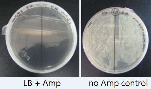
| + | | [[Image:NYMU_urea_poor_growth.jpg|400px]] | ||
| + | || | ||
| + | * 09/12, Ming-Han | ||
| + | * Ligation and/or transformation of ureR and ureDp failed again. Newly arrived DNA Ligase and its buffer are used. | ||
| + | * Next step: | ||
| + | ** Check the properties of all the materials used: | ||
| + | *** What antibiotic resistance contained in the vectors? --Ampicillin, Kanamycin, Chloramphenicol, or none!? | ||
| + | *** Produce new and fresh "ES" and "XP" digested vectors in the meanwhile. | ||
| + | *** Did the PCR products have enzyme cutting sites? | ||
| + | ** Look over all details of our design. | ||
| + | *** Were the restriction enzymes chosen correctly? | ||
| + | *** Were the inserts and vectors obtained reasonably? | ||
| + | *** Were the ratios of insert to vector reasonable? | ||
| + | ** Operate again using the hand of another. | ||
| + | *** Misoperation? | ||
| + | |- | ||
| + | | -- | ||
| + | || | ||
| + | * 09/18-21 | ||
| + | * Digest E0240 vector by EcoRI & SpeI in 2 steps; use gel electrophoresis to check each enzyme works. | ||
| + | * Digest PCR products (ureR & ureDp) by EcoRI & SpeI in 2 steps and check each enzyme works. | ||
| + | * Purify these digested DNA fragments. | ||
| + | * Ligate these DNA fragments in room temperature or 4 degrees Celsius | ||
| + | ** DO NOT ligate in 37 degrees Celsius, because the sticky end are too short to anneal in such high temperature. | ||
| + | ** Use newly arrived ligase. | ||
| + | ** Try more different insert-to-vector ratios. | ||
| + | * Transform them into competent cells. | ||
| + | * Select and amplify colonies expected. | ||
| + | * Get plasmids needed. | ||
| + | |- | ||
| + | | | ||
| + | || | ||
| + | * 09/21, Ming-Han | ||
| + | * PCR of ureR and ureDp failed; Both Pfu and Taq are failed. | ||
| + | * Using 4 different volumes of bacteria as template; see smear but no band. | ||
| + | * Positive control (E0240 plasmid) were amplified successfully. | ||
| + | * E0240 plsmid extraction secceeded. | ||
| + | |- | ||
| + | | | ||
| + | || | ||
| + | * 09/22, Ming-Han; Blent, Jesse, Tina et al. are consulted | ||
| + | * Gradient PCR and trying addtion of MgCl2; all are failed | ||
| + | * PCR program (Taq, gradient, 1500 bp, genomic DNA): | ||
| + | **STEP 1. 95, 3'00" | ||
| + | **STEP 2. 95, 1'00" | ||
| + | **STEP 3. 45~55, 1'00" | ||
| + | **STEP 4. 68, 3'00", go to step 2, 29 repeats | ||
| + | **STEP 5. 68, 5'00" | ||
| + | **STEP 6. 4, hold. | ||
| + | |- | ||
| + | | [[Image:NYMU_urea_plasmid1012_labeled.png|400px]] | ||
| + | || | ||
| + | * 10/11, Ming-Han | ||
| + | * Check transformation | ||
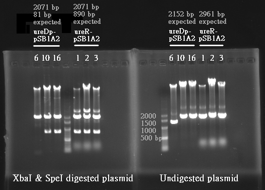
| + | * ureDp-pSB1A2 (left 3) and ureR-pSB1A2 (right 3) digested by XbaI and SpeI | ||
| + | * what are the >>2000bp bands? | ||
| + | |- | ||
| + | | [[Image:NYMU_urea_plasmid1014.png|400px]] | ||
| + | || | ||
| + | * 10/13, Ming-Han | ||
| + | * Check transformation | ||
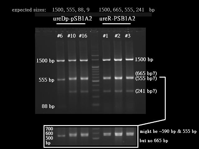
| + | * ureDp-pSB1A2 (left 3) and ureR-pSB1A2 (right 3) digested by RsaI and NotI | ||
| + | * Expected sizes: | ||
| + | ** ureDp-pSB1A2 (left 3) = 1500, 555, 88, 9 | ||
| + | ** ureR-pSB1A2 (right 3) = 1500, 665, 555, 241 | ||
| + | * Real sizes: | ||
| + | ** ureDp-pSB1A2 (left 1) #6 = 1500, 555, 88 | ||
| + | ** ureDp-pSB1A2 (left 2)#10 = 1500, 555, 280(what?) | ||
| + | ** ureDp-pSB1A2 (left 3)#16 = 1500, 555, 280(what?) | ||
| + | ** ureR-pSB1A2 (right 3) #1 = 1500, 555, 280(what?) | ||
| + | ** ureR-pSB1A2 (right 2) #2 = 1500, 555, 280(what?) | ||
| + | ** ureR-pSB1A2 (right 1) #3 = 1500, 555, 280(what?) | ||
| + | |- | ||
| + | |||
| + | |} | ||
Revision as of 16:52, 29 October 2008
| Home | Project Overview: | pH Sensor | Attachment | Time Regulation | Waste Removal | Experiments and Parts | About Us |
Contents |
Why should urea be removed
Physiologically, urea is synthesized from ammonia and carbon dioxide. The elimination of urea means getting rid of nitrogenous waste from the body. In healthy individuals, blood plasma are filtrated by kidney and the waste removed by urination; in patients with renal failure, such waste are accumulated in blood. These patients have to get renal dialysis treatment to remove the waste, or they would suffer from hepatic failure and encephalopathy, which is worse. For these reasons, urea should be removed by a healthy kidney or by medical treatment.
However, the renal dialysis treatment knocks down the quality of life because of the time spent being treated and equipment portage (or patient transportation). If urea can be removed by the enterobacteria (bacteria in intestines, such as E. coli or our BacToKidney), a capsule or an yogurt-like drug may help reduce the frequency of get renal dialyzed and increase the quality of life. If so, the next half of the patients' lifespan may be more fun.
How do we try to remove urea
Absorption Method--Urea Transporter (chosen method)
- Urea may be absorbed by the micro dialysis machine (or be uptaken by the engineered bacteria), and this micro machine may be eliminated with stool passage.
- Urea transporter would be expressed only if both pH sensor and urea sensor are turned on:
- When the device is turned on by pH sensor, ureR, the pH sensor is expressed. The ureR protein may bind with urea molecule to form a complex. This ureR-urea complex may activate a downstream promoter, ureDp, and then urea transporter will be expressed.
- In this circuit design, urea sensor turns not only urea transporter on but also guanidine transporter on at the same time. Guanidine transporter is integrated into this circuit because the concentratoin of guanidine in blood may be elevated after an rising of urea consentration.
Adsorption Method
- Urea remover 1: use albumin to adsorb urea on the surface of bacteria
- [http://www.ncbi.nlm.nih.gov/entrez/viewer.fcgival=NC_000004.10&from=74488870&to=74505996&dopt=gb human serum albumin] + [http://www.ncbi.nlm.nih.gov/Structure/cdd/wrpsb.cgi?seqinput=YP_001724652.1 E.coli TM domain]
- Urea remover 2: use urease to bind urea on the surface of bacteria
- [http://www.ncbi.nlm.nih.gov/sites/entrezDb=gene&Cmd=retrieve&dopt=full_report&list_uids=4099022&log$=databasead&logdbfrom=nuccore H.pylori urease (1)] [http://www.ncbi.nlm.nih.gov/sites/entrez?Db=gene&Cmd=retrieve&dopt=full_report&list_uids=900171&log$=databasead&logdbfrom=nuccore (2)] [http://www.ncbi.nlm.nih.gov/sites/entrez?Db=gene&Cmd=retrieve&dopt=full_report&list_uids=890414&log$=databasead&logdbfrom=nuccore (3)] + [http://www.ncbi.nlm.nih.gov/Structure/cdd/wrpsb.cgi?seqinput=YP_001724652.1 E.coli TM domain]
- Urea remover 3: "Lactobaccilize" the bacteria; produce more lactic acid and adsorb urea on the bacterial capsule in low pH level
- [http://www.ncbi.nlm.nih.gov/sites/entrezDb=gene&Cmd=retrieve&dopt=full_report&list_uids=944834&log$=databasead&logdbfrom=nuccore E.coli aceE, aceF, lpdA]
How to measure the concentration of urea
Urease method
Urea + H2O --(urease)--> 2NH4+ + CO2
NH4+ + alpha-KG + NADH --(GLDH)--> Glutamate + NAD+ + H2O
The change of NADH concentration reflects the urea concentration in the solution.
Absorbance of NADH = A340nm - A405nm
- use an in vitro diagnostic kit
- [http://www.fbc.com.tw/j2fbs/fbc_tw/medical/24-pdf/BUN_dream%201.0.pdf Formosa Biomedical Technology] Blood Urea Nitrogen diagnostic kit
- or undertaken by clinical laboratories
- [http://www.ucl.com.tw/WebMaster/?section=92 UCL (Union Clinical Laboratory)] A test for urea nitrogen costs NTD$60 (as private patient without health insurance)
Diacetyl monoxime method:
Diacetyl Monoxime + Urea --> Diazine (yellow)
- [http://www.searo.who.int/en/Section10/Section17/Section53/Section481_1754.htm WHO SOP]
- [http://www.clinchem.org/cgi/content/abstract/15/5/393 The Automated Thiosemicarbazide-Diacetyl Monoxime Method for Plasma Urea, Clinical Chemistry 15: 393-396, 1969]
 "
"

