Minnesota/24 June 2008
From 2008.igem.org
| Back to Notebook Home | |
| Go to Previous Day (June 23) | Go to Next Day (June 25) |
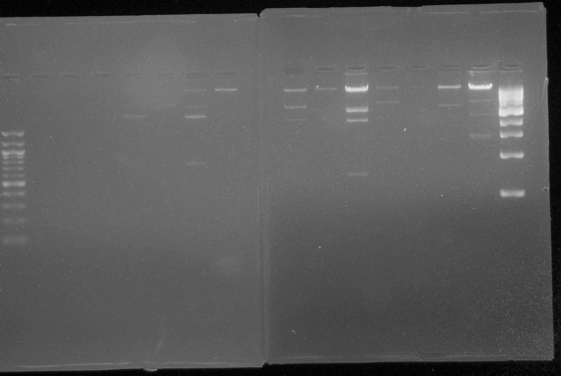
| 1. Gel Electrophoresis: Used this technique to show plasmid DNA sequence. Materials: |
| a. 50 uL of 1% agarose gel |
| b. Buffer TAE |
| c. One gram of 1% agarose per 100 uL of TAE |
| d. Ethidium bromide (intercalating agent) |
| Problem encountered: electrophoretic gels with 1% agarose had deficient wells |
| Solution: add 0.5 grams more of agarose to the 100uL of TAE buffer |
| 2. Plating from 6-23-08 transformations again. | ||||||||||||||||||||||||||||||||||||||||||||||||||||||||||||||||||||||||||
| a. Since plating of the 6-23 transformations provided no colonies for parts 15-18, the remaining cells from those transformations were replated. 75 uL of cell culture was spread on each of two plates for each culture; plates contained LB media and the corresponding antibiotic. A metal spreading tool was used to spread the culture suspension on the plates, and this was sterilized between each sample by dipping it in 100% ethanol (EtOH) and flaming it. 75 uL cell culture was pipetted on, and spread around plate. | ||||||||||||||||||||||||||||||||||||||||||||||||||||||||||||||||||||||||||
| b. Plates were placed at 37C in an incubator and allowed to grow overnight. | ||||||||||||||||||||||||||||||||||||||||||||||||||||||||||||||||||||||||||
3. Sequencing primers ordered on 6-20-08 were picked up. All primers were diluted to mircomoles according to the following additions of sterile H20:
|
 "
"
