Team:NTU-Singapore/Parts/Fluorescence Microscope Image
From 2008.igem.org
|
Images and Videos from the Characterization Lab
pLacI-GFP part:BBa_J04430 found to express RFP instead
The below pictures were taken after the BBa_J04430 was transformed into Top 10 competent cells. They clearly showed, either under normal light or UV light, the cells appears red or glow red respectively. This is contrary to what was expected, as we would expect it to glow green under UV light. This problem was highlighted to the iGEM Headquarters. In order to solve this problem the pLac promoter (R0010) was ligated with the GFP (E0040), allowing us to proceed further with the characterization experiments.
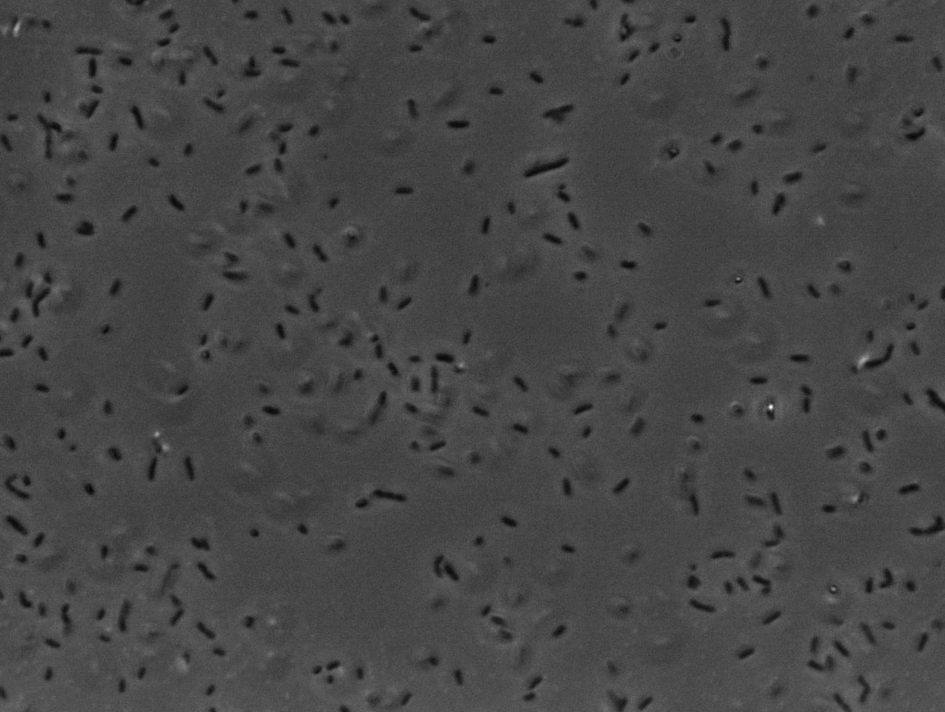
Image results obtained from fluorescence microscope
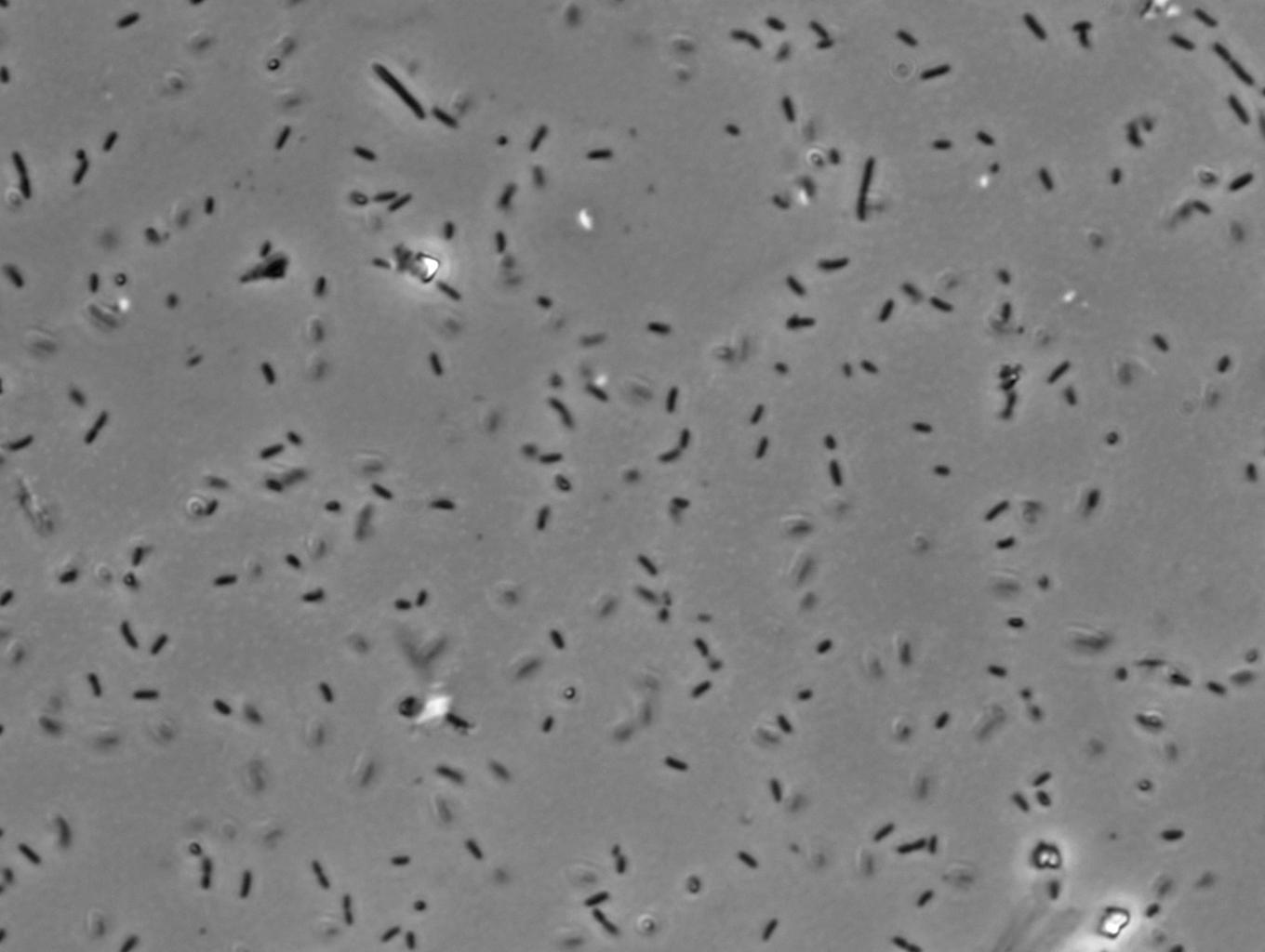
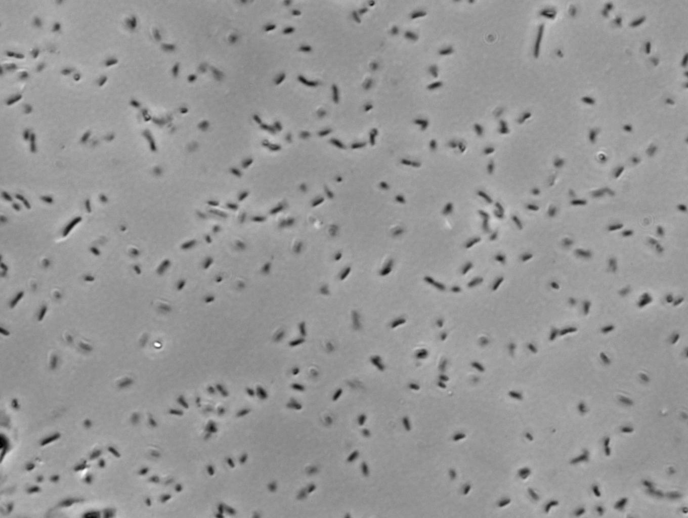
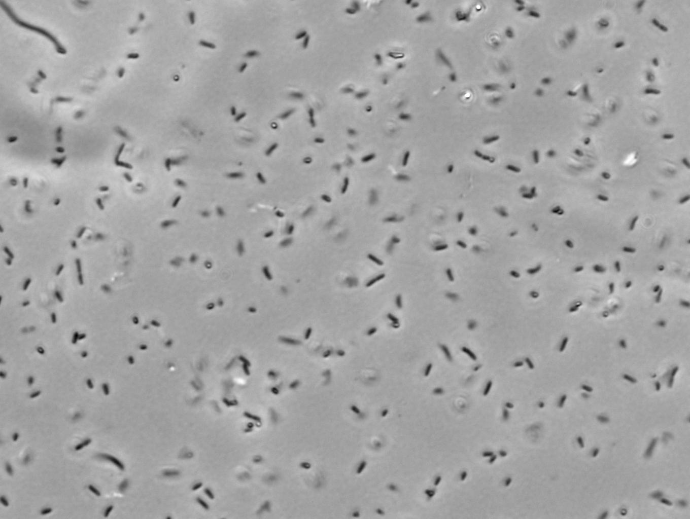
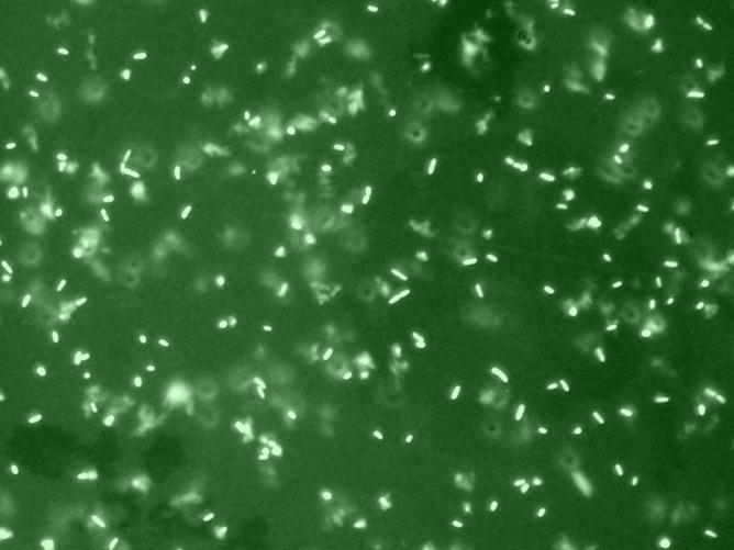
Below are Images obtained from the microscope of the various standards (Low, Medium, High & Standard) that iGEM Headquarters has provided for us in the [http://partsregistry.org/Measurement/SPU/Learn iGEM Newsletter].
Weak promoter [http://partsregistry.org/Part:BBa_J23150 BBa_J23150]
Medium promoter [http://partsregistry.org/Part:BBa_J23151 BBa_J23151]
Strong promoter [http://partsregistry.org/Part:BBa_J23102 BBa_J23102]
Standard promoter with GFP reporter [http://partsregistry.org/Part:BBa_I20260 BBa_I20260]
The above pictures showed the green fluorescence protein was produced by Top 10 competent cells that were taken under blue light excitation. These cells contained the different standard promoters with GFP and they have been allowed to grow to an OD of 1.2 before a snap shot was taken with the microscope.
Video obtained from fluorescence microscope
Video of E coli under normal white light
Video of E coli under blue light excitation
 "
"


