Team:University of Lethbridge/Notebook/Project1October
From 2008.igem.org
Back to The University of Lethbridge Main Notebook
Contents |
October 2, 2008
Munima, Christa, John
Objective: To set up chemotaxis spatial localization assay
Made and autoclaved media to make LB + amp plates to be used in the assay.
Recipe:
- Peptone 5g
- Yeast Extract 2.5 g
- NaCl 5 g
- Agar (0.25 %) 1.25 g
- dH2O 500 mL
October 4, 2008
Christa
Objective: To set up chemotaxis spatail locatization assay
Media made on October 2 looked funny.
Poured 23 plates (LB + amp 100ug/mL) anyway. Stored in iGEM 4 C fridge.
October 6, 2008
Christa, Alix
Objective: Make more solutions for future chemical competency experiments.
Made 500 mL of 100 mM CaCl2 solution and 500 mL of 80mM MgCl2-20mM CaCl2 solution. They are stored on the counter under the iGEM glass cabinet in 1 L media bottles.
Selina
Objective: Create motility media stab tubes controls for motility assay.
Inoculated two 5 mL LB media tubes, one with E. coli K12 (wild-type), the other with Staphylococcus epidermis and let incubate at 37 C for ~8 hours.
Stabbed motility media (no theophylline) with subcultures of either cell type, using inoculating loops (teeny loop part, essentially a rod).
-Positive control: E. coli K12 (wild-type)
-Negative control: Staphylococcus epidermis
Incubated for 48 hours at 37 C.
Selina
Streaked the "LB+ amp chemically 'competent' cells transformed with pUC19 control plasmid" plate (Aug. 26) with wild-type E. coli to test for ampicillin presence.
Incubated overnight at 37 C.
October 7, 2008
Munima, John, Roxanne
Objective: Assess the motility media stab tubes after one day of incubation.

| 
|
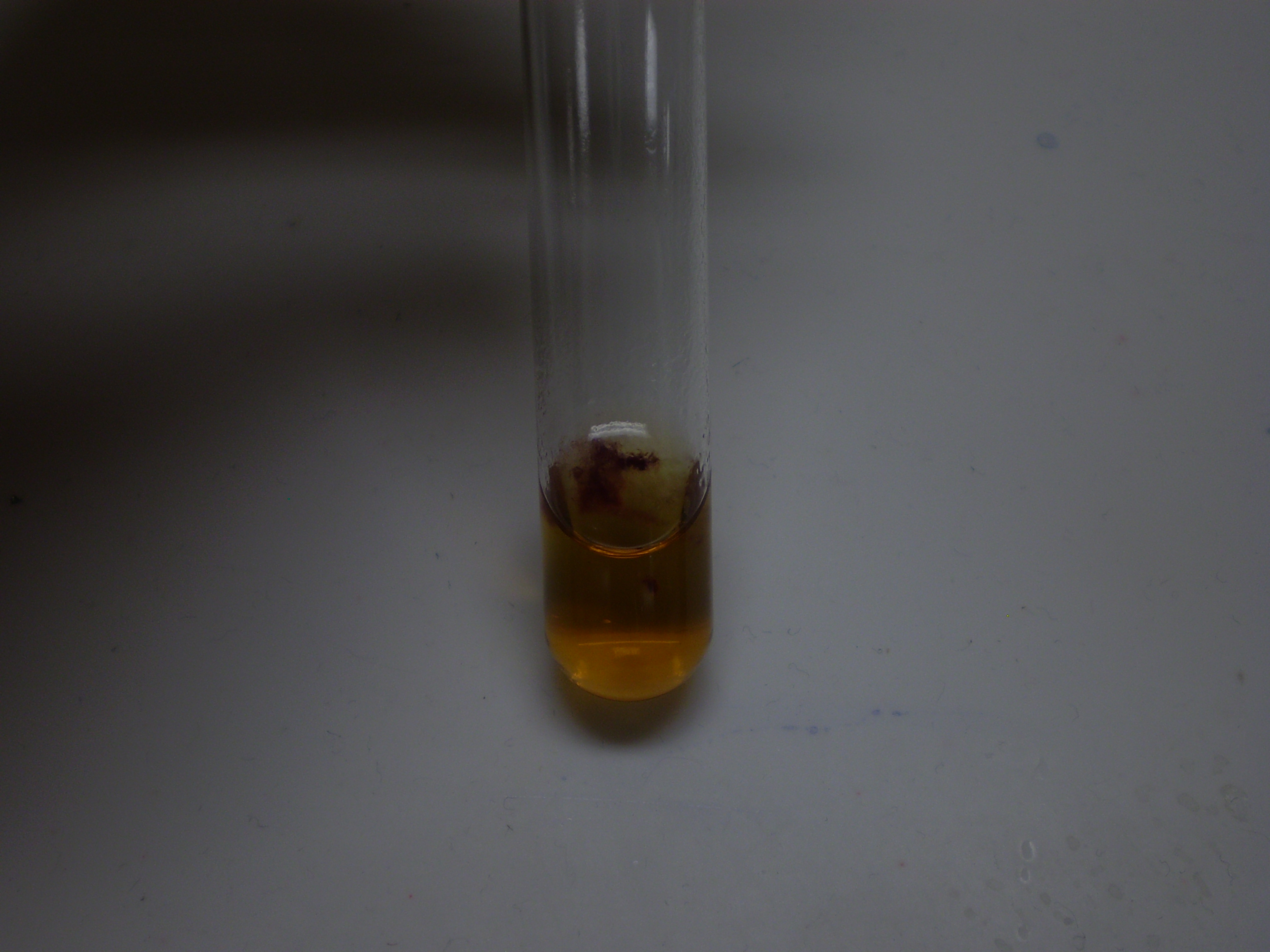
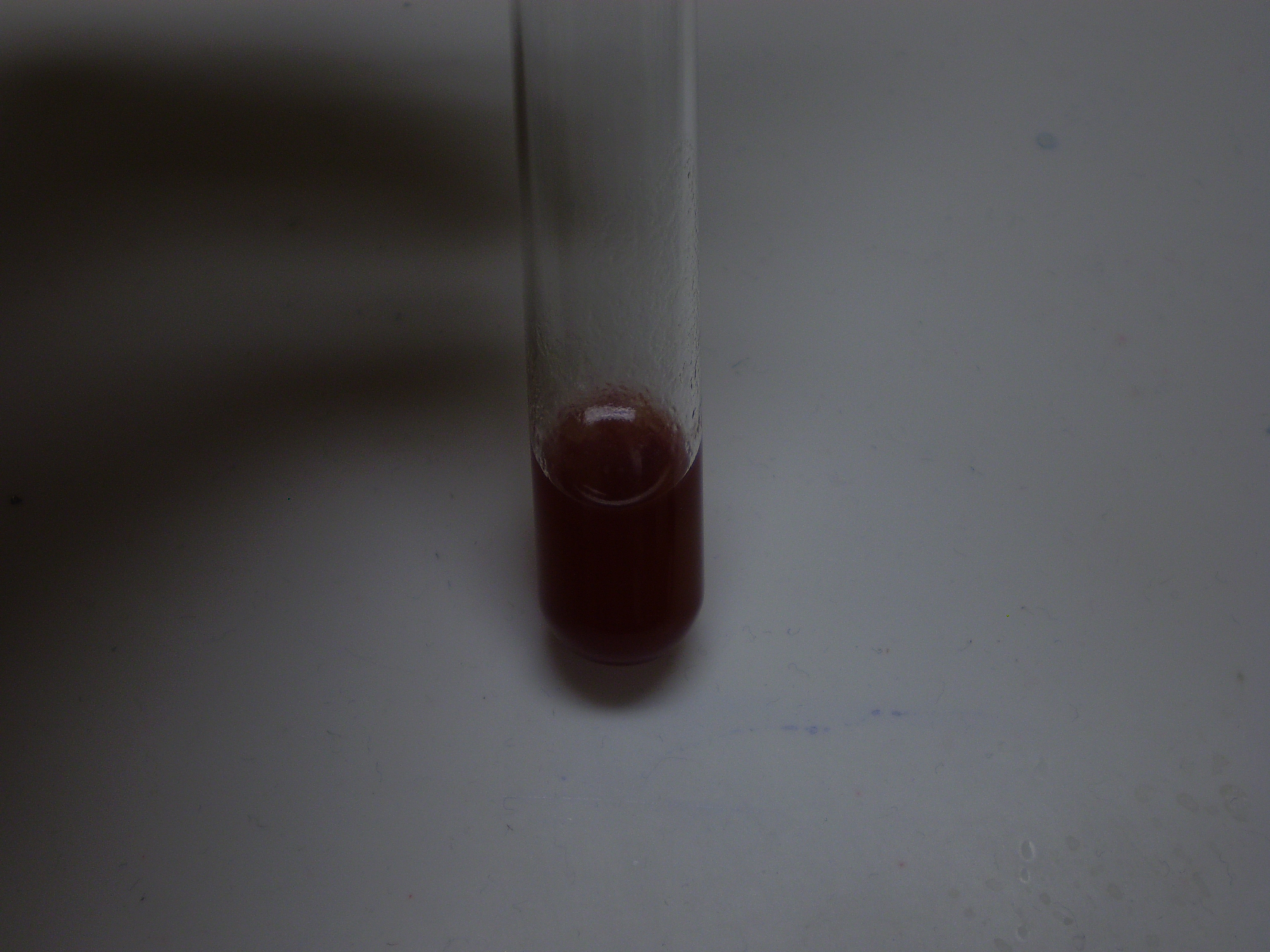
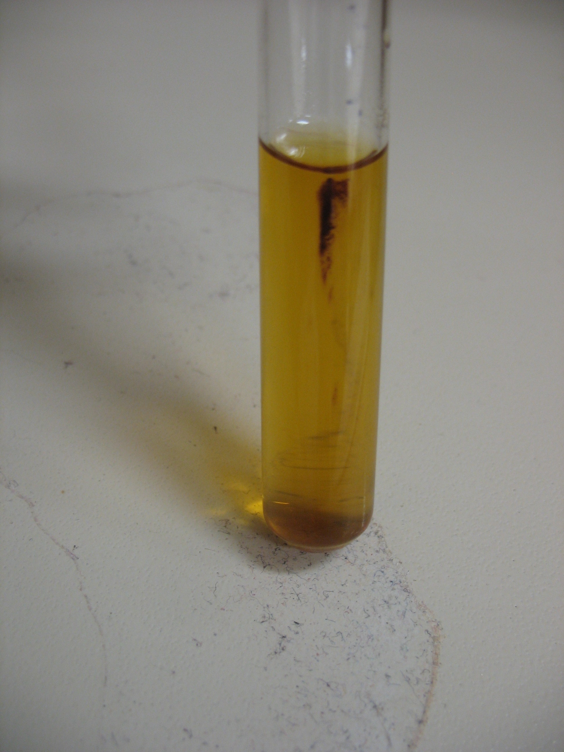
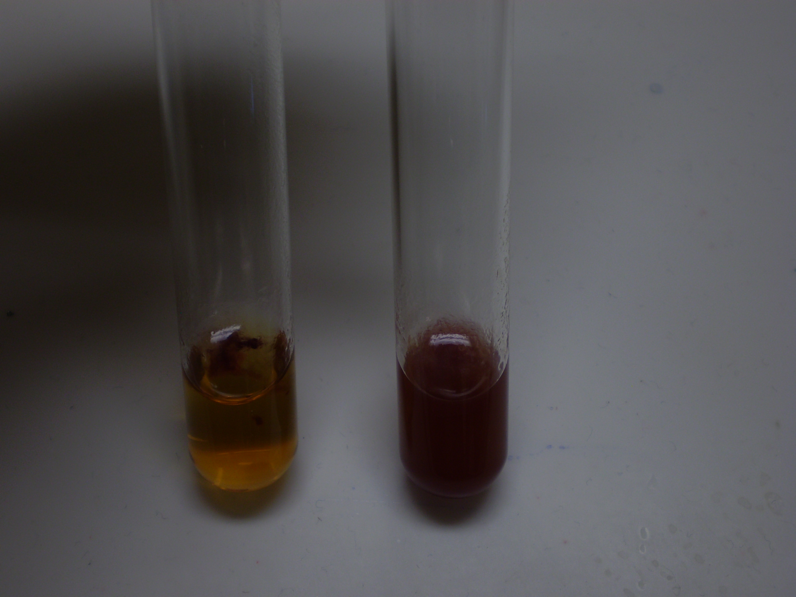
| Negative control: Staphylococcus epidermis | Positive control: K12 |
Side-by-side comparison:
Roxanne
Objective: Cut the CheZ gene with appropriate restriction endonucleases for insertion into a biobrick plasmid (pSB1A2) to be cut out of GFP complete system
-Restriction digested CheZ and GFP complete with XbaI and SpeI
-Needed pSB1A2 from GFP complete
-Gel extracting:
   -pLacI x 2
   -RFP x 2
   -TetR x 2
   -pSB1A2 x 2
   -CheZ
-Dephosphorylated pSB1A2 with Antarctic Phosphorylase to remove 5' Phosphates to ensure that
 self ligatioin does not occur
October 9, 2008
John
Objective: Attempt to make competent cells that can be used in future transformations.
Made 'chemically competent' cells of BL21 and RP1616. Stored in the bio stores -80 C freezer in 100 uL aliquots.
Christa, Munima
Objective: Assess the competency of the cells John prepared.
Did a transformation of BL21 and RP1616 with pUC19 (positive control) and pTopp using the protocol Jaden and Selina used on August 25, 2008.
Protocol (from Kothe Lab):
- Work Sterile! Open flame!
1. Thaw 20 uL of pre aliquoted cells (BL21 and RP1616) on ice. Competent cells are stored at -80 C.
  (Often cells are frozen as 50 uL aliquot - split under sterile conditions for two transformations).
  *throw away excess cells because they are no longer useful after being thawed*
2. Gently pipet 2.0 uL of DNA (pUC19 or pTopp) into the competent cells and invert
   *(protocol change; original said to pipet)
- ATTENTION: Never use more DNA than 10% of the volume of the competent cells.
  Otherwise the cells get destroyed by osmotic shock.
3. Mix the DNA into the cells by swirling the tip in the solution.
4. Incubate the cells on ice for 30 minutes.
5. Heat shock the cells in a water bath at 42 C for exactly 45 seconds.
6. Incubate fhe cells on ice for 1 minute.
7. Add 250 uL of sterile media (LB) to the cells and incubate at 37 C for 1 hour with shaking
   (tape microcentrifuge tubes in shaker incubator).
8. Label the LB plates on the outside perimeter with: your name, date, cell strain, plasmid,
   and volume plated.
9. Plate 100 uL and 50 uL on prewarmed LB plates containing the appropriate antibiotic (Ampicillin).
   For ligations: plate all 250 uL on 1 plate.
10. Leave plate for 10-15 min to soak the cell suspension into the agar.
11. Flip plate over (agar on top).
12. Incubate the plates in the 37 C oven overnight.
13. Keep the remaining solution in the 4 C fridge overnight until transformation has been confirmed.
Plates were: "comp" RP1616 + pUC19; "comp" RP1616 + pTopp; "comp" BL21 + pUC19; and "comp" BL21 + pTopp. 100 uL and 50 uL were plated for each onto LB + Amp plates.
October 10, 2008
Munima, Roxanne
Objective: Assess transformation results.
No growth on any of the plates. Forgot to do a 'transformation control' yesterday using DH5 alpha.
Roxanne will redo transformation today ("comp" RP1616 and BL21 cells with pUC19) using DH5alpha + pUC19 as a transformation control.
Protocol (from MAX Efficiency DH5alpha Competent Cells techinical sheet):
1. Thaw competent cells on wet ice (DH5alpha, BL21 and RP1616). Place required number of 17 x 100 mm
   polyproplene tubes (Falcon 2059) *(protocol change: we use 35 mL round bottom tubes) on ice.
2. Gently mix cells, then aliquot 100 uL of competent cells into chilled tubes.
3. Refreeze any unused cells in the dry ice/ethanol bath for 5 minutes before returned to the -80 C
   freezer. Do not use liquid nitrogen.
4. To determine the transformation efficiency, add 5 uL (50 pg) of pUC19 control DNA to one tube
   containing 100 uL competent cells. Move the pipette through the cells while dispensing. Gently
   tap to mix.
5. Extra step if using DNA from ligation reactions.
6. Incubate cells on ice for 30 minutes.
7. Heat-shock cells for 45 seconds in a 42 C water bath; do not shake.
8. Place on ice for 2 minutes.
9. Add 0.9 mL room temperature LB media (protocol change; original says SOC).
10. Shake at 225 rpm (37 C) for 1 hour.
11. Dilute the reaction containing the control plasmid DNA 1:100 with media. Spread 100 uL of
    this dilution on LB or YT plates with 100 ug/mL ampicillin.
12. Dilute the experimental reactions as necessary and spread 100 to 200 uL of this dilution
    as described in Step 11.
13. Incubate overnight at 37 C.
Plated 100 uL on LB + Amp plates.
October 21, 2008
Christa, John
-Transformed RP1616 with pTopp (x2), and BL21 with pUC19 (x2) as a control
- plated 100 uL (3 of each) on LB + amp
- stored in iGEM incubator overnight at 37 C
October 23, 2008
Christa
-Transformation results: one RP1616 + pTopp transformation successful
- Subcultured 3 colonies of RP1616 + pTopp in LB + amp liquid media, left overnight at 37 C in Weiden shaker incubator
- plated 50uL of RP1616 cells on LB plate, left this plate in the iGEM incubator at 37 C overnight
October 24, 2008
Christa & Munima
Inoculated a stab tube of motility media with RP1616 as a control; incubated at 37 C overnight. Streaked out RP1616 from Oct. 23 plates on LB.
October 25, 2008
Christa & Munima
Results of stab tube of RP1616 from October 24 yielded no growth. Did a stab tube of RP1616 + pTopp in motility media also. Incubated at 37 C overnight.
October 26, 2008
Christa
No growth observed in stab tube of RP1616 +pTopp.
October 27, 2008
Christa & Munima
Stabbed 0, 0.25, 1mM [theophylline] of motility media with each of the following:
- RP1616
- RP1616 + pTopp
- K12
- RP1616 + GFP complete
October 28, 2008
Christa
Stabbed 0, 0.25 and 1 mM [theophylline] of motility media (+amp) of RP1616 + pTopp. Incubated overnight at 37 C.
October 29, 2008
Christa
Motility media results:
- RP1616 +pTopp (+amp) are still growing (Oct. 28/08)
- RP1616 + pTopp (Oct. 27/08)- 0 mM [theophylline] growth mostly on top, with a little growth down the tube; photo below |- RP1616 + pTopp (Oct. 27/08)- 0.25 mM [theo] growth mostly on top, with a little growth down the tube; photo below |
- RP1616 + pTopp (Oct. 27/08)- 1 mM [theo] no growth observed.
- K12- motility media 0 mM [theo] growth observed; photo below |- K12- no growth observed in 0.25 mM and 1 mM [theo]
- RP1616- motility media 0 mM [theo] growth observed- cells lack motility- lines of cells where stabbed; photo below |- RP1616- 0.25 mM [theo] growth observed at the top and down the stab tube; photo below |
- RP1616- 1 mM [theo] growth observed - lines of cells where stabbed, no growth on top =cells lacking motility; photo below |

- RP1616 + GFP- 0 mM [theo] growth observed on the top of tube; photo below |- RP1616 + GFP- 0.25 mM [theo] growth observed on the top of the stab tube; photo below |
- RP1616 + GFP- 1 mM [theo] growth observed on the top and a little down the stab tube; photo below |

 "
"
