Team:University of Sheffield/Eva
From 2008.igem.org
| Introduction | Our project | Modelling | Parts | Lab Books | Calendar |
|---|
Contents |
Eva's Notebook
Management
Along with two other team members,we were actually running the first iGEM project within the University of Sheffield, even though this University has nearly the best Molecular Biology and Biotechnology Department in the UK.
Sponsorship
What is more we managed to collect material sponsorship as well as financial contribution worth of about £10 000. This includes free lab space and chemical materials in the Sheffield Bioincubator, a newly built commercial business and research facility, also free primers and CqsS gene synthesis worth more than £1000 from the IDTDNA comercial company. Finally, financial support towards traveling and living expenses from MBB department, Prof. R. Poole, Biofusion, iCHEME .
Biobrick Characterisation
The initial idea was to insert the GFP under the pgaABCD operon controlled by small regulatory RNA - CsrA. Once the CsrA inhibition of this operon is releaved under the sensing event of the fusion kinase, the GFP would be expressed. What is more, the project was to have the GFP with an LVA tag (AANDENYALAA). This means the GFP would have a tag targeted by proteases and would be degraded within 45 minutes once there is no more stimulation. An idea to include degradable GFP was taken from the Andersen, J.B et al. (1998) New Unstable Variants of Green Fluorescent Protein for Studies of Transient Gene Expression in Bacteria. Applied and Environmental Microbiology
Introduction
However, this plan was based on achieving a successful gene knock-out, which we failed to achieve. Nonetheless, simultaneously we were trying to carry out a characterization of a GFP-LVA biobrick. This particular Biobrick would be the best choice for our idea, as it would make the sensing bacteria re-usable within about 45 minutes after the triggering of the fusion kinase stops. Also, the Biobrick present in the booklet is under lac promoter, which means that we could vary the IPTG concentration to have the optimal GFP expression rate.
Schedule
- August 2008 - the transformation was carried out with CaCl2 chemically - competent DH5-alpha strains using the transformation method described in Sambrock.
- The transformation was very "weak", meaning that there was only one colony growing after transformation. It might have been due to increased kanamycin concentration used on the plates (50ugrams/ml instead of 30 ugrams/ml) as well as short recovery time (the cells were incubated at 37 degrees Celcius only for 30 minutes) .
- The transformant was replated and the fluorescence measurement was carried out using Tecan high-flow cytometer.
- Protocol used for characterization :
- 1.Grow 5ml of overnight cultures of DH5-alpha cells containing the plasmid with GFP-LVA.
- 2.Resuspend the overnight cultures in 50 ml of LB medium until the OD600 reaches about 0.6.
- 3.Quickly add 0.2 mM of IPTG to the medium and plate that into 96 well plate.
- 4.Use the high flow cytometry machine TecanR to measure the fluorescence every 15 minutes for 8 hours (excitation wv – 485 nm, emission wv- 535 nm )(including shaking = aeration of the cultures)
Results
Even repeating the protocol for 5 times the results were consistent.
1.A control sample with LB + IPTG had more fluorescence than LB media with cultures (this is due to turbidity caused by the growth of cultures, which reduces the shining of LB) (Fluorescence varied : 47108 to 41136 units) 2.LB + DH5-alpha had a range of fluorescence from about 42 000 units decreasing to 32 770 units. 3.LB + DH5-alpha + IPTG (expected high levels of fluorescence) the fluorescence measurement showed from about 46 000 units decreasing to 41 700 units over 8 hours of measurement
The measurement was taken 7 times, varying the amount of IPTG added to the medium from (0.3 mM to 0.05 mM) this did not seem to affect the measurements in any noticeable way.
Further actions
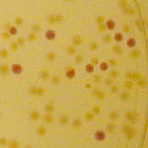
Together with this GFP – LVA characterization we decided to transform the cells using RFP (tested - working) and RFP-LVA (not characterised previously) and plating them directly on Amp + IPTG plates to straight-forwardly check for red transformant cells (Figure 1).
As it can be seen in the Figure 1 the RFP with no LVA tag was used by other iGEM teams. The transformed cells can be easily visualized under UV light in UV box as well as are visible under naked eye.
- The transformation with RFP and RFP-LVA was carried out on the 15th October
- The DNA content was checked before the actual procedure. The spectrophotometer indicated 91 ng/ul and 121 ng/ ul for RFP-LVA and RFP respectively
- The transformation was unsuccessful even the pre-checked chemically competent DH5-alpha cells were used, the positive controls worked and as a last attempt, the transformation was made by a Ph.D. student.
Conclusion
Unfortunately, the step that seemed to be the easiest one - transformation failed even after excessive attempts. A lot of troubleshooting was done, however nothing seemed to let us figure out what was the problem.
Once again:
- the transformation was carried out not only by us but also by an experienced Ph.D. student
- the cells and media used was carefully checked, and the same stock of cells seems to be working for positive controls
- even though the protocols used were not from the booklet, variation seemed to be too little to actually affect the success of the transformation. What is more, we used three various protocols for our experiment - none of them seemed to be successful
We are coming to the conclusion that there might have been something faulty with the booklet we received.
Leaflet Production
This was my second responsibility.
Software
Scribus, an open source desktop publishing program was selected as being most suitable for this task.
Initial Design Work
This is still a work in progress.
 "
"


