Team:Rice University/Notebook/14 June 2008
From 2008.igem.org
(Difference between revisions)
StevensonT (Talk | contribs) |
StevensonT (Talk | contribs) |
||
| Line 5: | Line 5: | ||
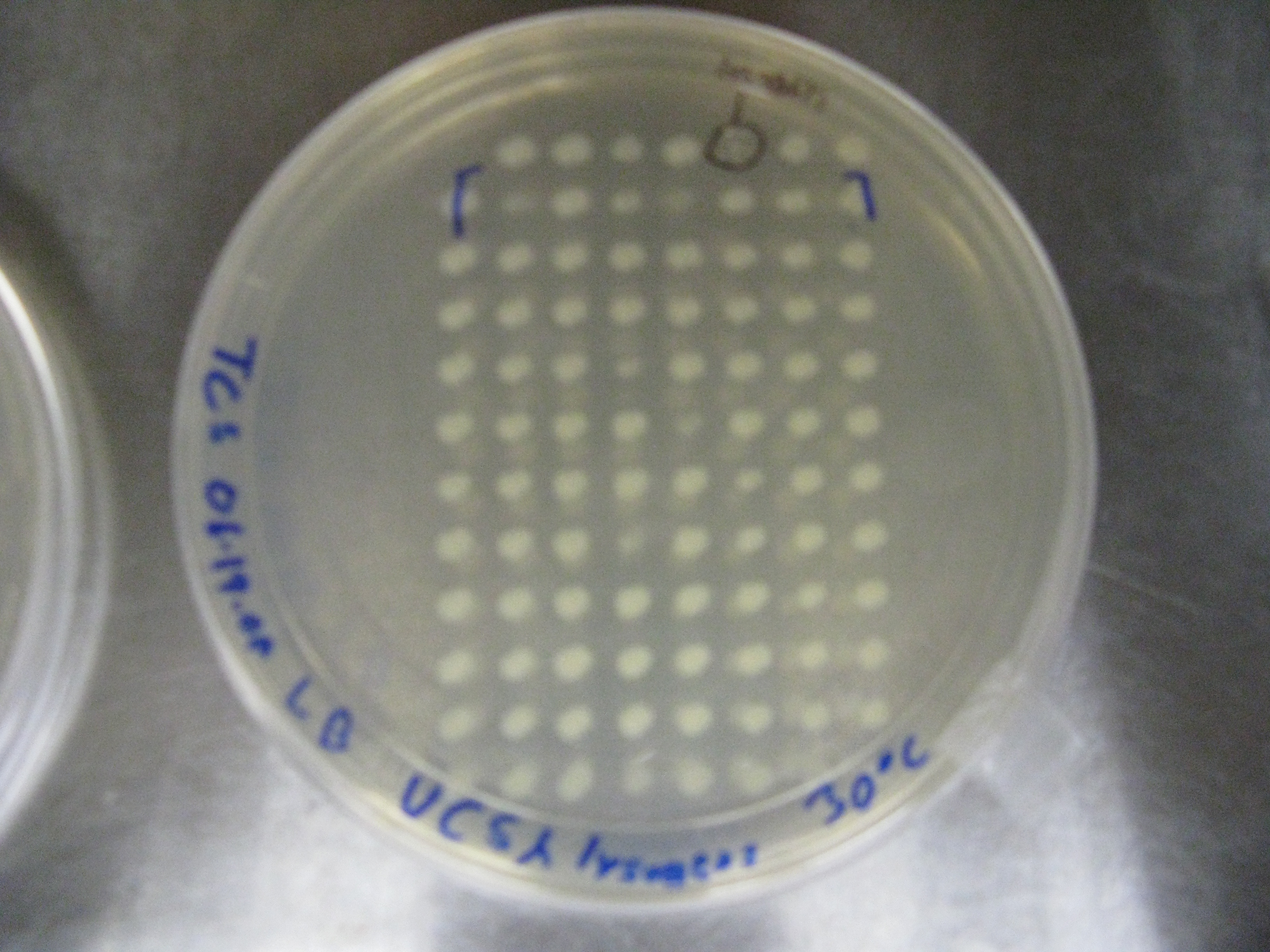
**'''Result'''-both plates showed bacterial growth at all but one spot after 24h. The one spot appears to be a lysogen. | **'''Result'''-both plates showed bacterial growth at all but one spot after 24h. The one spot appears to be a lysogen. | ||
| - | [[image:20080614_30*C_VCS_lysogen.jpg|300px| | + | [[image:20080614_30*C_VCS_lysogen.jpg|300px|center]] [[image:20080614_42*C_VCS_lysogen.jpg|300px|center]] |
| - | <BR><BR><BR><BR> | + | <BR><BR><BR><BR><BR><BR><BR><BR><BR> |
{| style="color:#1b2c8a;background-color:#0c6;" cellpadding="3" cellspacing="1" border="1" bordercolor="#fff" width="62%" align="center" | {| style="color:#1b2c8a;background-color:#0c6;" cellpadding="3" cellspacing="1" border="1" bordercolor="#fff" width="62%" align="center" | ||
Revision as of 20:13, 25 June 2008
Saturday 14 June
- Taylor Stevenson
- Phage infected VCS257 colonies cultured in 96 deep-well plate were spotted onto two LB plates using a 2uL plate replicator. Both plates were incubated O/N, one at 30*C and one at 42*C (temp is high enough to denature the CI repressor, causing any lysogen to become lytic).
- Result-both plates showed bacterial growth at all but one spot after 24h. The one spot appears to be a lysogen.
| Home | The Team | The Project | Parts Submitted to the Registry | Modeling | Notebook |
|---|
 "
"

