|
Home
The Team
Project Report
Parts
Modeling
Notebook
Safety
CoLABoration
|
Introduction:
Besides the project with the artificial receptor system, it was also tried to activate t-cells (B12.7.5) and b-cells (j558lδmmb1nfleck) with the NIP-linked DNA-origami. Both cell-types have a anti NIP Fab-fragment linked to their receptor. During these tries many problems were faced which could emerge as obstacles in the main project, the artificial receptor which is expressed by 293t-cells.
TThe first problem was the media. The cells were normally kept in Ringer solution during the first measurements which contains too little Mg2+ to keep the origamis stable. So the Mg2+ tolerance of the cells had to be tested in the hope the origamis would be stable if we add Mg2+ to the buffer.
Additionally the 293t-cells were tested on their tolerace to TA-buffer in which the origamis are stored.
Since the binding between Fab-fragment and NIP is essential for coupling of the receotors, Alexa 488 linked origamis with and without NIP were given to the cells to detect the binding and the fluorescence was visualized with a LSM.
In order to test a simple transfection method for the 293t cells, Ca2+ precipitation with a lac z gene was done and the β-galactosidase was detected with an ONPG test. To test the functionality of the transfectionvector and the CMV-promotor in it, CFP and YFP was cloned behind the promotor and the plasmid was brought into the 293t cells with the same method.
Materials and methods:
Mg2+ tolerance tests
T-cells:
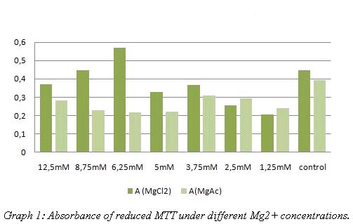
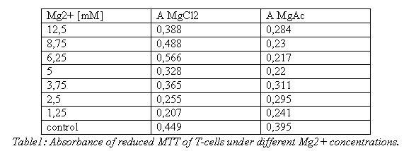
To test the Mg2+ tolerance of the T-cells, 800µl RPMI medium was mixed with 100µl cellsuspension and 100µl MgCl2/MgAc Solution with different concentrations and given to a 24-well plate. Because a qualitative result seemed to be sufficient, seeding the cells was done without counting them before. 3 days later the cells of each well were spun down, the supernatant was discarded and the cells were resolved in 200µl new RPMI medium. Additionally 50µl MTT was added to each sample. After 4h of incubation at 37°C the cells were spun down again and after discarding the medium the pellet was resolved in 400µl DMSO and 50µl Soerensens’ reagent. The reduced blue MTT was detected in the photometer at 570nm.
B-cells:
B-cells of an 10ml dish were spun down and resolved in 9ml Ringer (+12,5mM MgAc). After incubation for 45min the cells were spun down again and resolved in 1ml PBS. 5µl of the suspension was mixed with 45µl Trypan blue and the cells were counted in a “Neubauer cell chamber”. Since the dye can’t penetrate cells with active inner membrane potential, only dead cells got stained with this method.
293t-cells:
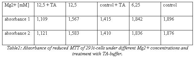
The cells were scraped off an 10ml dish, spun down and resolved in 10ml new DMEM medium. 500µl of this suspension was given in each plate of a 6-well plate containing 4500µl DMEM medium with different concentrations of Mg2+. 3 days later the media of 3 wells was sucked off and the cells were washed in PBS, then TA-buffer was given to these wells. After 1h the TA-buffer was removed, the cells of all dishes were washed in PBS and 2ml new DMEM medium plus 500µl MTT was added. After incubation for 3,5h at 37°C the cells were scraped off the wells and spun down at 13000 rpm for 5min. Then the pellet was resolved in 4ml DMSO and 500µl Soerensens’ reagent. Detection took place at 570nm.
Ca2+ precipitation
In order to transfect the 293T-cells, the amount of cells per ml was determined with the "Neubauer cell chamber" To transfect cells with 1µg DNA in each well of a 6-well plate, you need about 6*10E4 cells per well, so 20µl of the suspension was given in each well and 2ml DMEM was added. The next day, the cells were washed in PBS and new DMEM medium was added. 1/2h later 1µg DNA was mixed with 25µl CaCl2 and 224µl H20 dest. The mixture was given on ice for 20min and after that 250µl BBS (2x) was given to it. After 2 min the 500µl were given to the cells.
To prove the success of the lac z transfection an ONPG-assay was done. After 48h one part of the cells was harvested by washing them in PBS and scraping them off. Then the cells were centrifuged at 13000rpm for 2 min and the PBS was replaced by 500µl lysisbuffer (1x). Incubation took place at -80°C for 20min. After thawing the solution was vortexed, spun down and the supernatant was frozen at -20°C. The same procedure was done with the rest of the cells one day later (68h). Then 20µl of each lysate was given to 130µl reactionbuffer (incl. ONPG) letting the mixture incubate for 1h at 37°C. Measurement was done using the ELISA-reader at 405nm.
The detection of YFP took place 1 day later under a microscope with YFP filter.
Detection of the receptor-NIP linkage
To test the linkage between origamis and t-cells/b-cells, 15µl cell suspension in Ringer (12,5mM Mg2+) or TA-buffer (12,Italic text5mM Mg2+) was mixed with 15µl of origamis on a IBIDI µ-Slide. The linkage was regarded using a LSM.
Results:
Mg2+ Tolerance tests:
T-cells:
B-cells:
Counting stained and unstained B-cells brought following result:
Dead cells : 1
Living cells: 44
Total cell number: 45
293t-cells:
Ca2+ precipitation
Transfection of the trasfectionvector+cmv+YFP:
The transfected cells showed fluorescence by excitation of 510-520nm while the untransfected remained dark at this wavelength
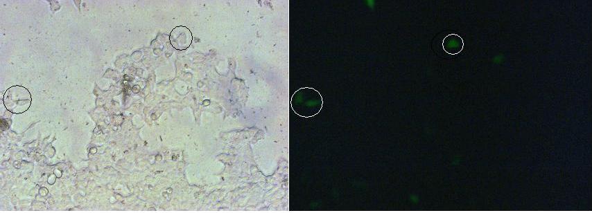
Picture1:Transfected cells without (left) and with YFP-filter (right)
ONPG-assay:

Table3: Absorbance of o-Nitrophenol produced by the β-galactosidase
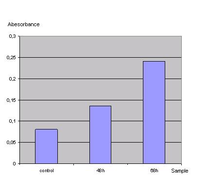
Graph2: Absorbance of o-Nitrophenol produced by the β-galactosidase
Control was done with untransfected cells using the same procedure.
|
 "
"


