Team:Rice University/Notebook/14 June 2008
From 2008.igem.org
Revision as of 15:43, 27 June 2008 by DavidOuyang (Talk | contribs)
Saturday 14 June
- Taylor Stevenson
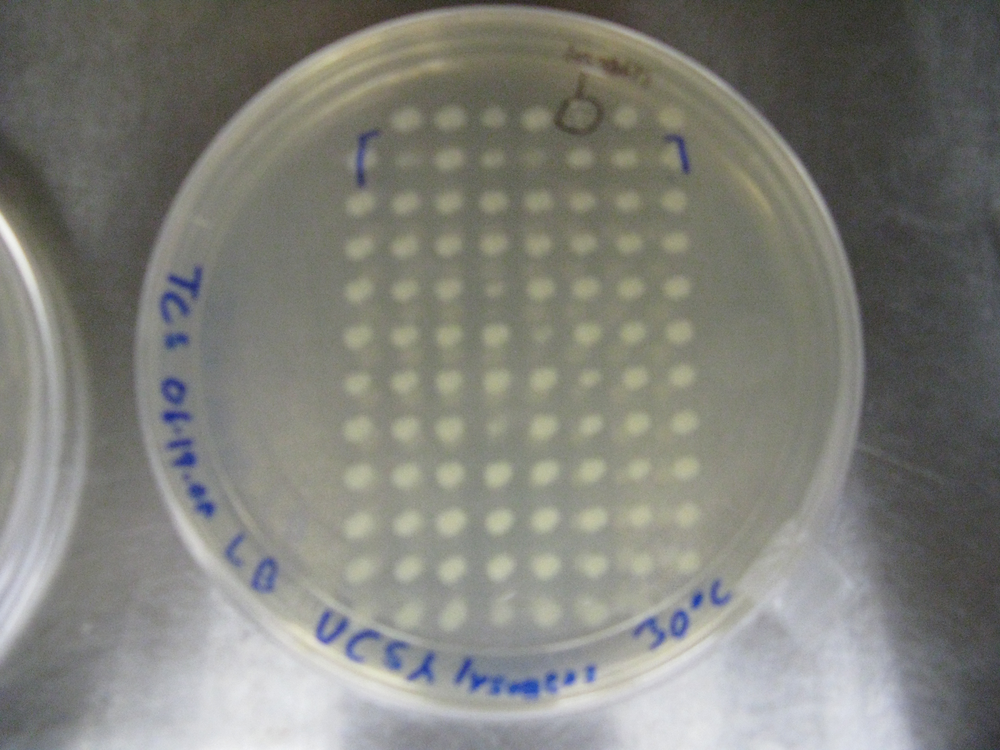
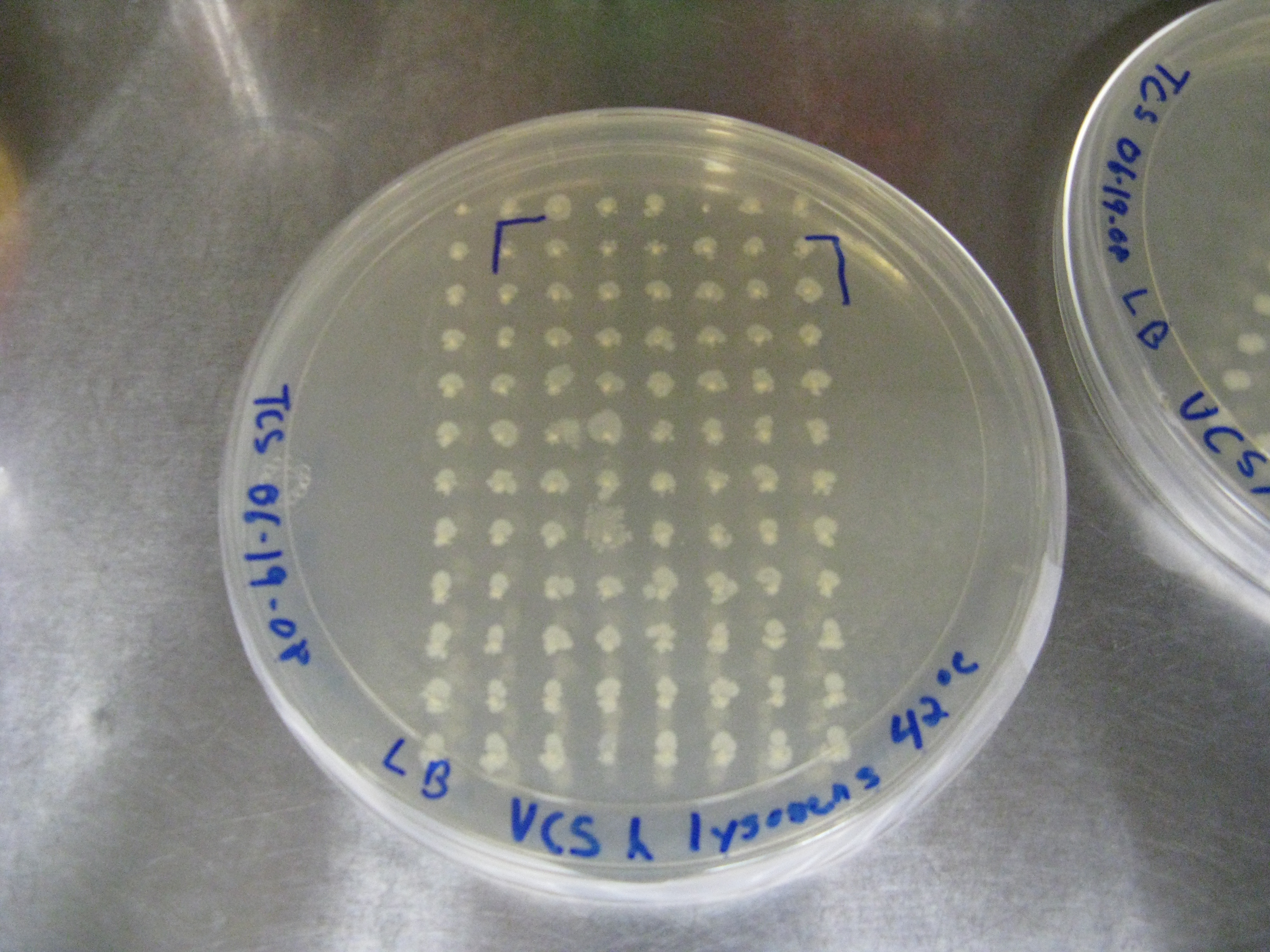
- Phage infected VCS257 colonies cultured in 96 deep-well plate were spotted onto two LB plates using a 2uL plate replicator. Both plates were incubated O/N, one at 30*C and one at 42*C (temp is high enough to denature the CI repressor, causing any lysogen to become lytic).
- Result-both plates showed bacterial growth at all but one spot after 24h. The one spot appears to be a lysogen.
The upper left spot on both plates is a negative control used to gauge aseptic technique. The only colony that appears to have grown @ 30*C, but not at 42*C was the third from the right at the top. This colony will be further screened for presence of a lysogen.
- David Ouyang
- PCR amplified the side tail fiber region (stf) and excision/integration segment (attp).
- Ran gel - looks good, sent out for sequencing.
| Home | The Team | The Project | Parts Submitted to the Registry | Modeling | Notebook |
|---|
 "
"

