Team:Chiba/Sender experiments/Senders(JW1908) T9002(JW1908)
From 2008.igem.org
(→Reaction temparature:37°C) |
(→Reaction temparature:30°C) |
||
| Line 50: | Line 50: | ||
===Reaction temparature:30°C=== | ===Reaction temparature:30°C=== | ||
====Sender culture:500μL,Receiver culture:500μL==== | ====Sender culture:500μL,Receiver culture:500μL==== | ||
| - | [[Image:Chiba_talks_JW1908_30_RS1_01.gif|thumb|left|'''Fig.5''' <br>E.coli strain,Senders:JW1908,BBa_T9002:JW1908,30°C]] | + | [[Image:Chiba_talks_JW1908_30_RS1_01.gif|thumb|left|'''Fig.5''' <br>E.coli strain,Senders:JW1908,BBa_T9002:JW1908,30°C.All measurements are averages from three replicate cultures with error bars representing standard deviations.]] |
| - | [[Image:Chiba_talks_JW1908_30_RS1_02.gif|thumb|left|'''Fig.6''' <br>E.coli strain,Senders:JW1908,BBa_T9002:JW1908,30°C]] | + | [[Image:Chiba_talks_JW1908_30_RS1_02.gif|thumb|left|'''Fig.6''' <br>E.coli strain,Senders:JW1908,BBa_T9002:JW1908,30°C.All measurements are averages from three replicate cultures with error bars representing standard deviations.]] |
<br clear=all> | <br clear=all> | ||
| Line 58: | Line 58: | ||
====Sender culture:100μL,Receiver culture:1000μL==== | ====Sender culture:100μL,Receiver culture:1000μL==== | ||
| - | [[Image:Chiba_talks_JW1908_30_RS2_01.gif|thumb|left|'''Fig.7''' <br>E.coli strain,senders:JW1908,BBa_T9002:JW1908,30°C]] | + | [[Image:Chiba_talks_JW1908_30_RS2_01.gif|thumb|left|'''Fig.7''' <br>E.coli strain,senders:JW1908,BBa_T9002:JW1908,30°C.All measurements are averages from three replicate cultures with error bars representing standard deviations.]] |
| - | [[Image:Chiba_talks_JW1908_30_RS2_02.gif|thumb|left|'''Fig.8''' <br>E.coli strain,senders:JW1908,BBa_T9002:JW1908,30°C]] | + | [[Image:Chiba_talks_JW1908_30_RS2_02.gif|thumb|left|'''Fig.8''' <br>E.coli strain,senders:JW1908,BBa_T9002:JW1908,30°C.All measurements are averages from three replicate cultures with error bars representing standard deviations.]] |
<br clear=all> | <br clear=all> | ||
No significant difference in fluorescence intensity between the culture containing [http://partsregistry.org/Part:BBa_K084007 BBa_K084007](plac+RhlI) gene transformed cells and the culture containing [http://partsregistry.org/Part:BBa_K084008 BBa_K084008](plac+RhlI(LVA)) gene transformed cells.We thought that the rate of AHL synthesis by each autoinducer synthase was much faster than the rate of its degradation by protease. | No significant difference in fluorescence intensity between the culture containing [http://partsregistry.org/Part:BBa_K084007 BBa_K084007](plac+RhlI) gene transformed cells and the culture containing [http://partsregistry.org/Part:BBa_K084008 BBa_K084008](plac+RhlI(LVA)) gene transformed cells.We thought that the rate of AHL synthesis by each autoinducer synthase was much faster than the rate of its degradation by protease. | ||
====Sender culture:10μL,Receiver culture:1000μL==== | ====Sender culture:10μL,Receiver culture:1000μL==== | ||
| - | [[Image:Chiba_talks_JW1908_30_RS3_02.gif|thumb|left|'''Fig.9''' <br>E.coli strain,senders:JW1908,BBa_T9002:JW1908,30°C]] | + | [[Image:Chiba_talks_JW1908_30_RS3_02.gif|thumb|left|'''Fig.9''' <br>E.coli strain,senders:JW1908,BBa_T9002:JW1908,30°C.All measurements are averages from three replicate cultures with error bars representing standard deviations.]] |
<br clear=all> | <br clear=all> | ||
The final fluorescence intensity of the culture containing [http://partsregistry.org/Part:BBa_K084008 BBa_K084008] transformed cells which express LVA-tagged RhlI protein was lower than the culture containing cells which express untagged RhlI protein.It shows that LVA-tagged RhlI protein was degraded by LVA-specific protase.However,fluorescence intensity of both cultures increased at the same time.We thought it was because the AHL concentration was quickly reached the threshold concentration and cells began to express gfp.After 2 hours,fluorescence intensity started to increase. | The final fluorescence intensity of the culture containing [http://partsregistry.org/Part:BBa_K084008 BBa_K084008] transformed cells which express LVA-tagged RhlI protein was lower than the culture containing cells which express untagged RhlI protein.It shows that LVA-tagged RhlI protein was degraded by LVA-specific protase.However,fluorescence intensity of both cultures increased at the same time.We thought it was because the AHL concentration was quickly reached the threshold concentration and cells began to express gfp.After 2 hours,fluorescence intensity started to increase. | ||
Revision as of 08:45, 30 October 2008
| Home | The Team | The Project | Parts Submitted to the Registry | Reference | Notebook | Acknowledgements |
|---|
Results and Discussion
Reaction temparature:37°C,09/12
Sender culture:1000μL,Receiver culture:1000μL
Sender culture:100μL,Receiver culture:1000μL
- Fig. 1,Fig. 2
Green fluorescence intensity didn't increase in the culture containing BBa_K0840010(plac+CinI+LVA).We thought BBa_K084010 didn't work properly or LuxR gene didn't interact with signal molecules synthesized by BBa_K084010. Others responded at the same time(4 hours after induction).Final fluorescence intensity differd.
Reaction temparature:37°C
Sender culture:500μLm,Receiver culture:500μL
- Fig. 3
Response time and final fluorescence intensity showed no significant difference. We thought AHL concentration was quickly reached the threshold concentration.The fluorescence intensity increased as gfp maturured.
- Fig. 4
No significant difference in fluorescence intensity between the culture containing BBa_K084007(plac+RhlI) gene transformed cells and the culture containing BBa_K084008(plac+RhlI(LVA)) gene transformed cells.We thought that the rate of AHL synthesis by each autoinducer synthase was much faster than the rate of its degradation by protease.
Reaction temparature:30°C
Sender culture:500μL,Receiver culture:500μL
- Fig. 6
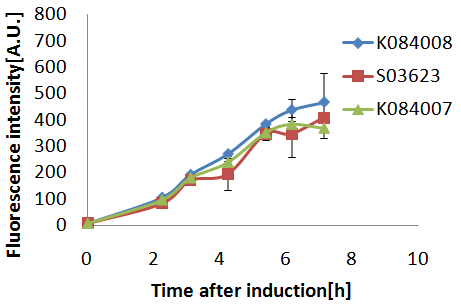
No significant difference in fluorescence intensity between the culture containing BBa_K084007(plac+RhlI) gene transformed cells and the culture containing BBa_K084008(plac+RhlI(LVA)) gene transformed cells.We thought that the rate of AHL synthesis by each autoinducer synthase was much faster than the rate of its degradation by protease.
Sender culture:100μL,Receiver culture:1000μL
No significant difference in fluorescence intensity between the culture containing BBa_K084007(plac+RhlI) gene transformed cells and the culture containing BBa_K084008(plac+RhlI(LVA)) gene transformed cells.We thought that the rate of AHL synthesis by each autoinducer synthase was much faster than the rate of its degradation by protease.
Sender culture:10μL,Receiver culture:1000μL
The final fluorescence intensity of the culture containing BBa_K084008 transformed cells which express LVA-tagged RhlI protein was lower than the culture containing cells which express untagged RhlI protein.It shows that LVA-tagged RhlI protein was degraded by LVA-specific protase.However,fluorescence intensity of both cultures increased at the same time.We thought it was because the AHL concentration was quickly reached the threshold concentration and cells began to express gfp.After 2 hours,fluorescence intensity started to increase.
>Back to Sender experiment and result
 "
"


