Team:Bologna/Results
From 2008.igem.org
Rita.morini (Talk | contribs) (→UV Spectrum) |
Rita.morini (Talk | contribs) (→UV Spectrum) |
||
| Line 25: | Line 25: | ||
<br> | <br> | ||
[[Image:spettro.jpg|center]] | [[Image:spettro.jpg|center]] | ||
| + | <br> | ||
| + | The killing spectrum of UV light coincides with the peak absorbance of DNA (near 260nm) and is caused by the dimerization of two adjacent thymines. The best studied transcriptional response to DNA damage is the SOS response [Friedberg et al., 1995; Walker, 1996]. | ||
| + | This systems can be divided into two class: the SOS Photoreactivation repair and the SOS triggered by RecA protein. The first uses the photolyase, a poorly expressed enzyme(encoded by genes phrA and phrB) which binds the pyrimidine dimers and uses the blue light to split them apart as the following picture shows. | ||
| + | [[Image:fosfoliasi.jpg|right]] | ||
Revision as of 08:29, 28 October 2008
| HOME | PROJECT | TEAM | SOFTWARE | MODELING | WET LAB | SUBMITTED PARTS | BIOSAFETY AND PROTOCOLS | RESULTS |
|---|
UV Spectrum
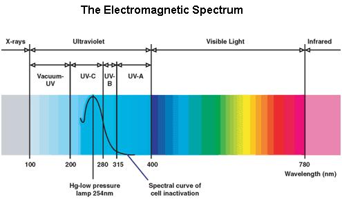
Ultraviolet is that part of electromagnetic radiation bounded by the lower wavelength extreme of the visible spectrum and the upper end of the X-ray radiation band. The spectral range of ultraviolet radiation is, by definition, between 100 and 400 nm and is invisible to human eyes. The UV spectrum is subdivided into three bands: UV-A (long-wave) from 315 to 400 nm, UV-B (mediumwave) from 280 to 315 nm, UV-C (short-wave) from 100 to 280 nm. A strong germicidal effect is provided by the radiation in the short-wave UV-C band.
The killing spectrum of UV light coincides with the peak absorbance of DNA (near 260nm) and is caused by the dimerization of two adjacent thymines. The best studied transcriptional response to DNA damage is the SOS response [Friedberg et al., 1995; Walker, 1996].
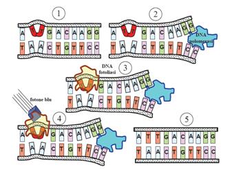
This systems can be divided into two class: the SOS Photoreactivation repair and the SOS triggered by RecA protein. The first uses the photolyase, a poorly expressed enzyme(encoded by genes phrA and phrB) which binds the pyrimidine dimers and uses the blue light to split them apart as the following picture shows.
 "
"


