Team:Freiburg Calcium Imaging
From 2008.igem.org
(Difference between revisions)
m |
|||
| Line 18: | Line 18: | ||
in RPMI (10% FCS), but the phenol red itself is an electron acceptor | in RPMI (10% FCS), but the phenol red itself is an electron acceptor | ||
and would disturb the measurement. Another complication was, that the | and would disturb the measurement. Another complication was, that the | ||
| - | Origami need a high | + | Origami need a high Mg<sup>2+</sup> concentration (12.5 mM), which stabilizes the |
DNA backbone, but low concentration of other bivalent cations, which | DNA backbone, but low concentration of other bivalent cations, which | ||
could disrupt the Origami. None of the common cell culture medium does | could disrupt the Origami. None of the common cell culture medium does | ||
| Line 24: | Line 24: | ||
the stability of the Origami in different media were tested (see | the stability of the Origami in different media were tested (see | ||
DNA-Origami). On the other hand we also had to test if our cells | DNA-Origami). On the other hand we also had to test if our cells | ||
| - | survive 12.5 mM | + | survive 12.5 mM Mg<sup>2+</sup>, which we tested with an MTT-Assay. <br> |
As explained before, we also wanted to use the Origami to activate | As explained before, we also wanted to use the Origami to activate | ||
T-cell receptors (TCR) by clustering. For this experiment we measured | T-cell receptors (TCR) by clustering. For this experiment we measured | ||
| Line 45: | Line 45: | ||
<h1>Material and Methods</h1> | <h1>Material and Methods</h1> | ||
<br> | <br> | ||
| - | <h2>Cell stability in the presence of | + | <h2>Cell stability in the presence of Mg<sup>2+</sup> measured by MTT-Assay</h2> |
| - | To test the | + | To test the Mg<sup>2+</sup> tolerance of the T-cells (cell line |
B.12.7.5), 100 µl cellsuspension was mixed with 800 µl RPMI | B.12.7.5), 100 µl cellsuspension was mixed with 800 µl RPMI | ||
medium and 100 µl MgCl2 or MgAc, respectively | medium and 100 µl MgCl2 or MgAc, respectively | ||
| - | containing various concentrations of | + | containing various concentrations of Mg<sup>2+</sup> in a 24-well plate. 3 days |
later cells of each well were spun down, the supernatant was | later cells of each well were spun down, the supernatant was | ||
discarded and the cells were resuspended in 200 µl new RPMI medium. 50 | discarded and the cells were resuspended in 200 µl new RPMI medium. 50 | ||
| Line 65: | Line 65: | ||
<br> | <br> | ||
| - | 293T cells were scraped off an 10 ml dish, spun down and resolved in 10 ml new DMEM medium. 500 µl of this suspension was given in each plate of a 6-well plate containing 4500 µl DMEM medium with different concentrations of | + | 293T cells were scraped off an 10 ml dish, spun down and resolved in 10 ml new DMEM medium. 500 µl of this suspension was given in each plate of a 6-well plate containing 4500 µl DMEM medium with different concentrations of Mg<sup>2+</sup>. 3 days later the media of 3 wells was sucked off and the cells were washed in PBS, then TA-buffer (Tris-Acetat buffer) was given to these wells. After 1h the TA-buffer was removed, the cells of all dishes were washed in PBS and 2ml new DMEM medium plus 500 µl MTT was added. After incubation for 3.5h at 37°C the cells were scraped off the wells and spun down at 13000 rpm for 5min. Then the pellet was resolved in 4 ml DMSO and 500 µl Soerensens’ reagent. Detection took place at 570 nm.<br> |
<br> | <br> | ||
<h2>Media</h2> | <h2>Media</h2> | ||
| Line 88: | Line 88: | ||
</ul> | </ul> | ||
<br> | <br> | ||
| - | Krebs-Ringer-Hepes (12.5 mM | + | Krebs-Ringer-Hepes (12.5 mM Mg<sup>2+</sup>):<br> |
<ul> | <ul> | ||
<li>NaCl (155 mM)</li> | <li>NaCl (155 mM)</li> | ||
| Line 110: | Line 110: | ||
<h2>Binding measurement</h2> | <h2>Binding measurement</h2> | ||
To test the binding between origamis and T-cells/B-cells 15 µl cell | To test the binding between origamis and T-cells/B-cells 15 µl cell | ||
| - | suspension in Ringer (12.5 mM | + | suspension in Ringer (12.5 mM Mg<sup>2+</sup>) or TA-buffer (12.5 mM Mg<sup>2+</sup>) was mixed |
with 15 µl of origamis on a µ-Slide (ibidi, µ-Slides 18 well-flat, Cat. | with 15 µl of origamis on a µ-Slide (ibidi, µ-Slides 18 well-flat, Cat. | ||
No: 81824). Those slides are coated with Poly-L-Lysine, which fixes the | No: 81824). Those slides are coated with Poly-L-Lysine, which fixes the | ||
| Line 117: | Line 117: | ||
<br> | <br> | ||
<br> | <br> | ||
| - | <h2> | + | <h2>Calcium<sup>2+</sup> measurement</h2> |
<br> | <br> | ||
| - | <h3> | + | <h3>Ca<sup>2+</sup> measurement with microscope</h3> |
By binding of ligands to a receptor at the cell surface the cell reacts | By binding of ligands to a receptor at the cell surface the cell reacts | ||
amongst others with a efflux of calcium ions from the ER into the | amongst others with a efflux of calcium ions from the ER into the | ||
| Line 125: | Line 125: | ||
quantify the concentration or rather the increase of calcium ions in the | quantify the concentration or rather the increase of calcium ions in the | ||
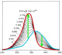
cytoplasm. Fura-2 is a fluorescent dye which change the quality | cytoplasm. Fura-2 is a fluorescent dye which change the quality | ||
| - | dependent on the | + | dependent on the Ca<sup>2+</sup> concentration. Fura-2AM (Fura-2-acetoxymethyl |
ester) is a membrane-permeable derivative of Fura-2 but after crossing | ester) is a membrane-permeable derivative of Fura-2 but after crossing | ||
the membrane the acetoxymethyl groups are removed by cellular esterases | the membrane the acetoxymethyl groups are removed by cellular esterases | ||
| Line 131: | Line 131: | ||
and 380 nm of light, and the ratio of the emissions at those | and 380 nm of light, and the ratio of the emissions at those | ||
wavelengths is directly correlated to the amount of intracellular | wavelengths is directly correlated to the amount of intracellular | ||
| - | calcium. Without | + | calcium. Without Ca<sup>2+</sup> the maximum emission results from excitation at |
| - | 365nm. With | + | 365nm. With Ca<sup>2+</sup> the maximum emission change to excitation at 340 nm and |
the emission decrease by extinction at 380 nm.<br> | the emission decrease by extinction at 380 nm.<br> | ||
So to measure properly it is necessary to alternate quickly between the | So to measure properly it is necessary to alternate quickly between the | ||
| Line 152: | Line 152: | ||
7. + 100 µl Ringer-Solution for resuspension<br> | 7. + 100 µl Ringer-Solution for resuspension<br> | ||
<br> | <br> | ||
| - | <h3> | + | <h3>Ca<sup>2+</sup> measurement with FACS</h3> |
Cells resuspended in medium with 1% serum were incubated with 5 μg/ml | Cells resuspended in medium with 1% serum were incubated with 5 μg/ml | ||
| - | of Indo-1, which is the | + | of Indo-1, which is the Ca<sup>2+</sup> complexing dye, and 0.5 μg/ml of |
pluronic F-127, which fasilitates dye uptake (both Molecular Probes) 45 | pluronic F-127, which fasilitates dye uptake (both Molecular Probes) 45 | ||
min at 37°C. After incubation, cells were distributed into to 1.5 ml | min at 37°C. After incubation, cells were distributed into to 1.5 ml | ||
eppendorf tubes and the washed with the medium we wanted to measure | eppendorf tubes and the washed with the medium we wanted to measure | ||
them. After washing, cells were resuspended in the according medium and | them. After washing, cells were resuspended in the according medium and | ||
| - | kept on ice. | + | kept on ice. Ca<sup>2+</sup> response was induced by addition of the indicated |
| - | stimulus 1 min after starting to record the ratio of | + | stimulus 1 min after starting to record the ratio of Ca<sup>2+</sup>-bound Indo-1 |
versus unbound Indo-1 with a LSRII fluorescence spectrometer (Becton | versus unbound Indo-1 with a LSRII fluorescence spectrometer (Becton | ||
Dickinson). Cells were measured for approximately 2min before putting | Dickinson). Cells were measured for approximately 2min before putting | ||
| Line 166: | Line 166: | ||
<br> | <br> | ||
<h1>Results and discussion</h1> | <h1>Results and discussion</h1> | ||
| - | <h2>Cell stability in the presence of | + | <h2>Cell stability in the presence of Mg<sup>2+</sup> measured by MTT-Assay</h2> |
'''T-cells:'''<br> | '''T-cells:'''<br> | ||
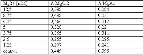
[[Image:Freiburg08tabellet-cellstability.JPG|Freiburg08tabellet-cellstability.JPG]]<br> | [[Image:Freiburg08tabellet-cellstability.JPG|Freiburg08tabellet-cellstability.JPG]]<br> | ||
| - | <small>Table 1: Absorbance of reduced MTT of T-cells with various | + | <small>Table 1: Absorbance of reduced MTT of T-cells with various Mg<sup>2+</sup> concentration</small><br> |
<br> | <br> | ||
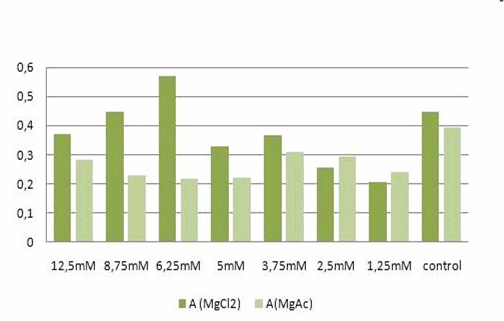
[[Image:TeamFreiburg2008-t-cellstability1.png]]<br> | [[Image:TeamFreiburg2008-t-cellstability1.png]]<br> | ||
| Line 182: | Line 182: | ||
'''293T-cells:'''<br> | '''293T-cells:'''<br> | ||
[[Image:TeamFreiburg2008_TABELLE-t-cellstability3.jpg ]]<br> | [[Image:TeamFreiburg2008_TABELLE-t-cellstability3.jpg ]]<br> | ||
| - | <small>Table 2: Absorbance of reduced MTT of 293T-cells with various | + | <small>Table 2: Absorbance of reduced MTT of 293T-cells with various Mg<sup>2+</sup> concentration and TA treatment</small><br> |
<br> | <br> | ||
The MTT assays and the trypan blue staining proofed the tolerance of | The MTT assays and the trypan blue staining proofed the tolerance of | ||
| - | the used cells towards a concentration up to 12.5 mM | + | the used cells towards a concentration up to 12.5 mM Mg<sup>2+</sup>. This is the |
exact concentration in which the origami are produced and stored. The | exact concentration in which the origami are produced and stored. The | ||
lower absorbance in the tests with TA could possibly come from the | lower absorbance in the tests with TA could possibly come from the | ||
| Line 192: | Line 192: | ||
cells might be sucked off with the TA.<br> | cells might be sucked off with the TA.<br> | ||
<br> | <br> | ||
| - | <h2> | + | <h2>Calcium<sup>2+</sup> measurement</h2> |
| - | <h3> | + | <h3>Ca<sup>2+</sup> measurement with FACS</h3> |
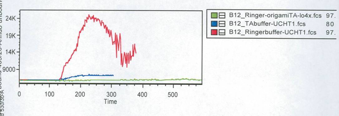
In this measurement we tried to activate T-Cells by clustering. | In this measurement we tried to activate T-Cells by clustering. | ||
Therefore we tested two different buffers, Krebs-Ringer-Hepes with | Therefore we tested two different buffers, Krebs-Ringer-Hepes with | ||
| - | 12.5 mM | + | 12.5 mM Mg<sup>2+</sup> buffer and TA with 12.5 mM Mg<sup>2+</sup>. As positive control we used |
UCHT1 (=anti-CD3), which can stimulate T-cells (Susana Minguet, Vol. | UCHT1 (=anti-CD3), which can stimulate T-cells (Susana Minguet, Vol. | ||
26, Page 43-54).<br> | 26, Page 43-54).<br> | ||
| Line 224: | Line 224: | ||
have to be carried out again.<br> | have to be carried out again.<br> | ||
<br> | <br> | ||
| - | <h3> | + | <h3>Ca<sup>2+</sup> measurement with microscope</h3> |
This measurement was also used to activate the T-cell receptors (TCR) | This measurement was also used to activate the T-cell receptors (TCR) | ||
by clustering. The TCR's were modified with a anti-NIP antibodies and | by clustering. The TCR's were modified with a anti-NIP antibodies and | ||
| Line 241: | Line 241: | ||
In contrast to the positive control (Pervanadat) which was working quit | In contrast to the positive control (Pervanadat) which was working quit | ||
well, our sample (DNA-origami with NIP) and the negative control | well, our sample (DNA-origami with NIP) and the negative control | ||
| - | (DNA-origami without NIP) did not show a significant | + | (DNA-origami without NIP) did not show a significant Ca<sup>2+</sup> efflux. There |
are two reasons which could be responsible that the cell answer to | are two reasons which could be responsible that the cell answer to | ||
origamis with and without NIP’s almost looks the same.<br> | origamis with and without NIP’s almost looks the same.<br> | ||
| Line 253: | Line 253: | ||
not blocked.<br> | not blocked.<br> | ||
<br> | <br> | ||
| - | In both cases the slow | + | In both cases the slow Ca<sup>2+</sup> efflux could result from the mechanical |
touch between the cells by adding the liquid with the probes.<br> | touch between the cells by adding the liquid with the probes.<br> | ||
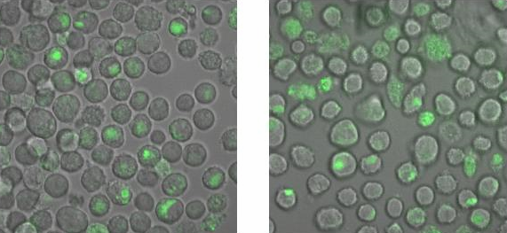
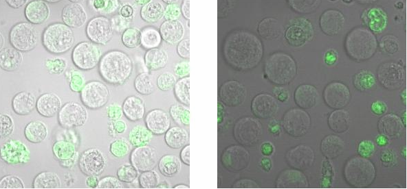
<h2>Binding measurement</h2><br> | <h2>Binding measurement</h2><br> | ||
| Line 260: | Line 260: | ||
[[Image:TeamFreiburg2008_Bindungsmessung_1.jpg|600 px]]<br> | [[Image:TeamFreiburg2008_Bindungsmessung_1.jpg|600 px]]<br> | ||
<small>Fig. 5: B-cells with NIP linked Origami (left) and without NIP (right) in 50% TA (Tris-Acetat) and 50% Krebs-Ringer-Hepes buffer, </small><br> | <small>Fig. 5: B-cells with NIP linked Origami (left) and without NIP (right) in 50% TA (Tris-Acetat) and 50% Krebs-Ringer-Hepes buffer, </small><br> | ||
| - | <small>both buffers contain 12.5 mM | + | <small>both buffers contain 12.5 mM Mg<sup>2+</sup> </small> |
<br> | <br> | ||
<br> | <br> | ||
Latest revision as of 00:58, 30 October 2008
 "
"

