|
← Yesterday ↓ Calendar ↑Tomorrow →
Digestion and ligation of the PCR we did yesterday
Yesterday we amplified flhD, flhC, flhDC with its promoter and E040 RBS+
Before the digestion, we have to determine the DNA concentration of the templates.
Measurement of DNA concentration
We used the biophotometer. We put 5 µL of template DNA in 95 µL of water. The Blank was made with 5 µL of EB Buffer (the one that was used for the elution of the DNA) in 95 µL of water.
| Template
| Concentration
(µg/µL)
|
PCR 130
E0240 RBS +
| 0.44
|
PCR 131
flhD RBS -
| 0.06
|
PCR 132
flhC RBS -
| 0.12
|
PCR 133
flhDC with promoter
| 0.08
|
MP 142
pSB3K3
| 0.04
|
MP 122
pSB1A2
| 0.2
|
Digestion
Protocol
| Digestion name
| Template DNA
| Enzymes
| Volume of DNA
|
| D 140
| PCR 130 - E0240 RBS +
| PstI
| 5 µL
|
| D 141
| PCR 131 - flhD rbs -
| EcoRI-SpeI
| 5 µL
|
| D 142
| PCR 132 - flhC rbs -
| EcoRI-SpeI
| 5 µL
|
| D 143
| PCR 133 - flhDC + prom
| EcoRI-SpeI
| 5 µL
|
| D 144
| MP 142 - pSB3K3
| EcoRI-SpeI
| 5 µL
|
| D 145
| MP122 - pSB1A2
| EcoRI-SpeI
| 5 µL
|
- X µL of Template DNA
- Buffer (n°2) 10X : 3µL
- BSA 100X : 0.3µL
- Pure water qsp 30 µL
- 1 µL of each enzyme
- Incubate during about 3h at 37°C, then 20 minutes at 65°C (to inactivate the enzymes).
- Exclusively for D 144 : 2 µL of Antarctic Phosphatase was added, 30 min at 37°C then 5 min at 65°C.
Results of the digestion
    For the digestion after PCR, an electrophoresis is useless : we can not separate DNA fragments that have
    10 bp of difference.

    Exclusively  for D 145 a gel extraction was made because there was two fragments, the standard protocol
    was used (the picture is missing because we hadn't the USB key).
    The DNA purification after gel extraction was done according the standard protocol.
Ligation
| Ligation name
| Insert name
| Volume of insert µL
| Vector name
| Volume of Vector µL
|
| L 139
| D 140 (E0240 RBS +)
| 3
| D 144 (pSB3K3)
| 3
|
| L 140
| D 141 (flhD)
| 3
| D 145 (pSB1A2)
| 3
|
| L 141
| D 142 (flhC)
| 3
| D 145 (pSB1A2)
| 3
|
| L 142
| D 143 (flhDC + promoter)
| 3
| D 145 (pSB1A2)
| 3
|
    REMARKS :

    Someone forgot to do the autoligation control, which is a key step in the evaluation of the ligation process.
Cloning on fliL promoter
Protocol
We used the Taq polymerase to amplify this promoter.
- Preparation of the template :
Resuspension of 1 colony E.coli K12 strain MG 1655 in 100µl of water.
1 µl O 124
1 µl O 125
1 µl template DNA
25 µL Quick Load Taq polymerase Mix
22 µL pure water
Result
We did an electrophoresis to check if our amplicon has the right size.
Actually, the electrophoresis did not show anything, not even the DNA ladder.
We decided to do again the electrophoresis tomorrow morning.
- Protocol (see # 3) Experiments done by QIAcube
| Name
| Ligation
| Biobricks
| Description
|
| MP144.1
| L128.1
| pFlgA-RFP
D132 (FI) - D136 (FV)
|  
|
| MP144.2
| L128.2
|
| MP144.3
| L128.3
|
| MP144.4
| L128.4
|
| MP145.1
| L129.1
| pFlgB-RFP
D133 (FI) - D136 (FV)
|  
|
| MP145.2
| L129.2
|
| MP145.6
| L129.6
|
| MP145.7
| L129.7
|
| MP146.1
| L130.1
| pFlhB-RFP
D134 (FI) - D136 (FV)
|  
|
| MP146.2
| L130.2
|
| MP146.7
| L130.7
|
| MP146.8
| L130.8
|
Glycerol Stocks
| Strain
| Ligation
| Biobricks
| Description
|
| S143.1
| L128.1
| pFlgA-RFP
D132 (FI) - D136 (FV)
|  
|
| S143.2
| L128.2
|
| S143.3
| L128.3
|
| S143.4
| L128.4
|
| S144.1
| L129.1
| pFlgB-RFP
D133 (FI) - D136 (FV)
|  
|
| S144.2
| L129.2
|
| S144.6
| L129.6
|
| S144.7
| L129.7
|
| S145.1
| L130.1
| pFlhB-RFP
D134 (FI) - D136 (FV)
|  
|
| S145.2
| L130.2
|
| S145.7
| L130.7
|
| S145.8
| L130.8
|
Results of the transformations we did yesterday
| Ligation name
| What's in it ?
| Number of colonies
|
| L 132
| flhDC (gene)
| 6
|
| L 133
| OmpR*
| 1
|
| L 134
| EnvZ*
| 11
|
| L 135
| pflgA
| 20
|
| L 136
| pflgB
| 100
|
| L 137
| pflhB
| 100
|
| L 138
| E0240
| 1
|
PCR screening of the transformations we did yesterday
Concentration of the MiniPreps
| Miniprep
| Concentration (µg/mL)
| Ratio 260/280
|
| MP144.1
| 127
| 1.67
|
| MP144.2
| 166
| 1.66
|
| MP144.3
| 163
| 1.57
|
| MP144.4
| 154
| 1.63
|
| MP145.1
| 136
| 1.67
|
| MP145.2
| 151
| 1.63
|
| MP145.6
| 163
| 1.67
|
| MP145.7
| 188
| 1.57
|
| MP146.1
| 298
| 1.70
|
| MP146.2
| 142
| 1.64
|
| MP146.7
| 170
| 1.52
|
| MP146.8
| 143
| 1.55
|
New PCR screening with the right primers
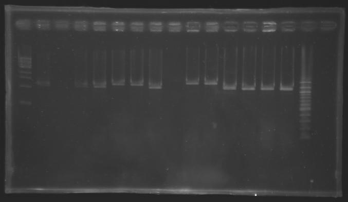
Transformants with pFlgA, pFlgB and pFlhB cloned into J61002 are analysed by PCR but this time with the right primers: VF2 (O18) and VR (O19).
- template: bacteria from glycerol stock
- positive control: J61002 (with ptet-mRFP)
- negative control: no template
Electrophoresis
- 1% agarose gel
- 10 µL of each sample loaded
| well n°
| 1
| 2
| 3
| 4
| 5
| 6
| 7
| 8
| 9
| 10
| 11
| 12
| 13
| 14
| 15
| 16
|
| sample
| 1 kb DNA ladder
| positive control
| negative control
| L128 (pFlgA)
| L129 (pFlgB)
| L130 (pFlhB)
| 100 bp DNA ladder
|
| clone n°
|
| 1
| 2
| 3
| 4
| 1
| 2
| 6
| 7
| 1
| 2
| 7
| 8
|
|
| red fluorescence
|
| yes
| yes
| no
| no
| yes
| yes
| no
| no
| yes
| no
| no
| no
|
|
| expected size
|
| 1161 bp
| 0 bp
| 1339 bp
| 1339 bp
| 1338 bp
|
|
| measured size
|
| 1,2 kb
| 0 kb
| 1,2 kb
| 1,2 kb
| 1,3 kb
| 1,3 kb
| 1,2 kb
| 0 kb
| 1,4 kb
| 1,4 kb
| 1,2 kb
| 1,2 kb
| 1,2 kb
| 1,2 kb
|
|
|
 "
"

